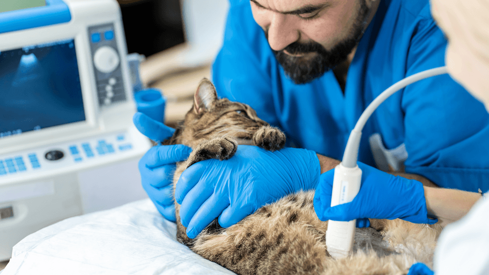
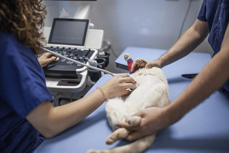
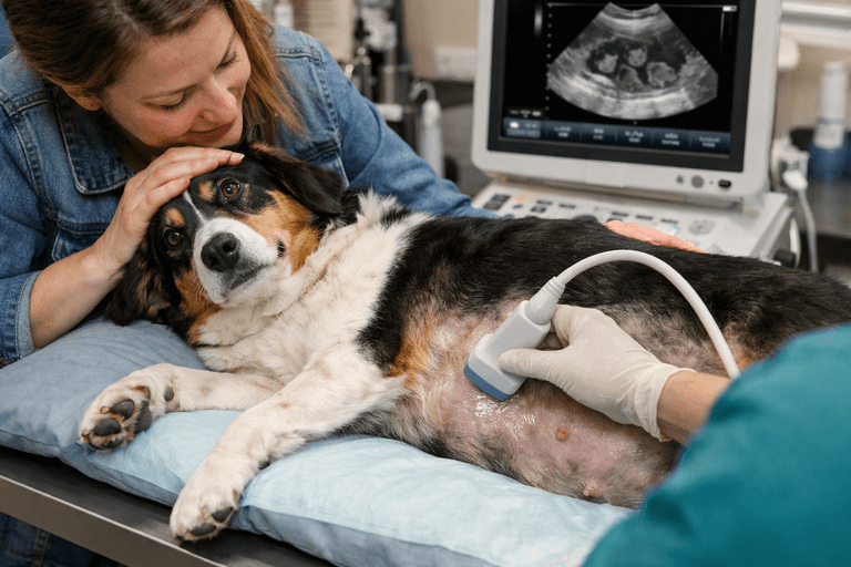
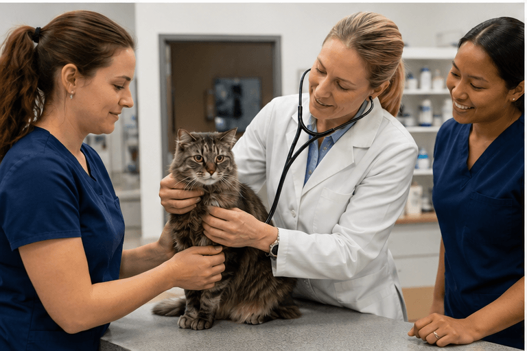
One of the most common things veterinarians identify during routine examinations is a heart murmur. For many pet owners, hearing the words “heart murmur” immediately creates anxiety. Some assume it automatically means heart failure. Others believe it is harmless and requires no further evaluation. In reality, a heart murmur is not a diagnosis by itself. A murmur simply means that abnormal or turbulent blood flow is being heard within the heart.The cause, severity, and long-term significance can vary tremendously from one patient to another. Some murmurs are relatively mild and may never progress into serious disease. Others may indicate structural heart abnormalities that require ongoing monitoring, medication, or adjustments in lifestyle and medical care. That is one reason veterinary echocardiography — also known as cardiac ultrasound or “echo” — has become such an important part of modern veterinary medicine. At Veterinary Diagnostic Centers, echocardiograms help veterinarians look beyond the sound of the murmur itself to evaluate what is actually happening inside the heart in real time. Advanced cardiac imaging allows us to assess heart valves, muscle thickness, blood flow, chamber size, and pumping function with far greater detail than physical exams or X-rays alone. For many pets, earlier cardiac evaluation helps support better long-term wellness, safer treatment planning, and more informed decision-making. What Exactly Is a Heart Murmur? A heart murmur is the sound created by turbulent blood flow within the heart or nearby blood vessels.During a physical examination, veterinarians listen for murmurs using a stethoscope. Murmurs are commonly described based on: Location Loudness Timing Intensity Associated heart rhythm abnormalities However, the sound of the murmur alone does not explain what structural problem may be causing it. This is where echocardiography becomes extremely valuable. An echocardiogram allows veterinarians to actually visualize the heart in motion and evaluate whether abnormalities are affecting: Heart valves Blood flow direction Chamber size Heart muscle thickness Pumping ability Pulmonary pressures Without an echocardiogram, it is often impossible to fully understand the significance of the murmur. Why Some Heart Murmurs Are More Serious Than Others Not all murmurs indicate severe disease. Some young puppies and kittens develop innocent murmurs that disappear as they mature.Other pets may have mild murmurs associated with relatively stable conditions that require only periodic monitoring. However, murmurs can also indicate significant cardiac disease involving: Degenerative valve disease Cardiomyopathy Congenital heart defects Pulmonary hypertension Dilated cardiomyopathy Valve malformations Heart enlargement The severity of the murmur does not always perfectly match the severity of the disease. Some pets with loud murmurs remain relatively stable for years. Others with softer murmurs may still have significant structural abnormalities internally. That is why echocardiography is considered the gold standard for evaluating cardiac disease in dogs and cats. What Is an Echocardiogram? An echocardiogram is a specialized ultrasound focused specifically on the heart. Unlike chest X-rays, which primarily show overall heart size and shape, echocardiographyallows veterinarians to evaluate the internal structures of the heart in real time. Cardiac ultrasound helps assess: Valve movement Chamber enlargement Blood flow patterns Wall thickness Pumping efficiency Pulmonary blood pressure Cardiac muscle disease Congenital abnormalities This detailed information helps veterinarians determine: Whether heart disease is present Which structures are affected Whether disease is mild or advanced Whether medication may be necessary How closely the condition should be monitored For many patients, echocardiography becomes one of the most important tools for long-term cardiac wellness planning. Common Heart Diseases Seen on Echocardiograms Degenerative Valve Disease in Dogs One of the most common cardiac diseases in dogs involves degeneration of the mitral valve. As the valve deteriorates over time, blood begins leaking backward through the heart, creating turbulence and eventually leading to heart enlargement. This condition is especially common in small-breed and aging dogs. Echocardiography helps veterinarians evaluate: Severity of valve leakage Degree of heart enlargement Pumping function Presence of congestive heart failure Pulmonary hypertension Monitoring progression early often allows veterinarians to begin treatment before advanced heart failure develops. Cardiomyopathy in Cats Cats commonly develop a very different form of heart disease known as cardiomyopathy. Instead of valve degeneration, the heart muscle itself becomes abnormally thickened. This thickening can interfere with normal blood flow and heart function. Unfortunately, many cats with cardiomyopathy show very subtle symptoms until disease becomes advanced. An echocardiogram helps veterinarians evaluate: Heart muscle thickness Chamber size Blood flow abnormalities Risk of clot formation Overall cardiac function Early identification is particularly important because some cats may appear outwardly healthy despite significant cardiac changes internally. Symptoms That May Indicate a Pet Needs an Echocardiogram Heart murmurs are one of the most common reasons veterinarians recommend an echocardiogram, but they are not the only reason. Additional symptoms that may prompt cardiac ultrasound include: Coughing Exercise intolerance Increased respiratory effort Weakness Collapse episodes Abnormal heart rhythms Fatigue Decreased stamina Rapid breathing Difficulty breathing In some pets, these symptoms may develop gradually and be mistaken for aging or reduced activity levels. Advanced cardiac imaging often helps determine whether the heart may be contributing to those changes. Certain Breeds Are More Predisposed to Heart Disease Some breeds are known to have significantly higher risk for cardiac disease.Examples include: Cavalier King Charles Spaniels Dobermans Boxers Maine Coon cats Ragdoll cats For predisposed breeds, echocardiography may sometimes detect disease earlier in the process before severe symptoms develop. Earlier detection often allows veterinarians to monitor progression more closely and intervene sooner when necessary. Why Echocardiograms Often Change Treatment Plans One of the most important aspects of veterinary diagnostics is that ruling out major disease can be just as valuable as confirming it. Even when an echocardiogram does not reveal severe disease, it still provides critical information that helps guide treatment decisions. Echocardiography may influence: Medication recommendations Anesthetic planning Surgical decisions Activity recommendations Long-term monitoring intervals Emergency risk assessment Prognosis discussions In some cases, cardiac ultrasound provides reassurance that a murmur is relatively mild. In others, it identifies advanced disease requiring more aggressive management. Both outcomes are extremely valuable for veterinarians and pet owners alike. Why Repeat Echocardiograms Are Often Necessary Heart disease is rarely static. Many cardiac conditions gradually progress over time, even in pets that initially appear stable. Repeat echocardiograms allow veterinarians to monitor: Valve progression Heart enlargement Changes in pumping function Pulmonary pressures Response to medication Disease progression This ongoing monitoring helps veterinarians adjust treatment plans appropriately as conditions evolve. Cardiac follow-up becomes especially important because some changes occur slowly and may not cause visible symptoms until disease becomes more advanced. Common Misconceptions About Echocardiograms Many pet owners feel nervous when cardiac ultrasound is recommended. Fortunately, most echocardiograms are far less stressful than owners expect. “My Pet Will Need Sedation” Most pets tolerate echocardiograms extremely well without sedation.In many cases, patients simply lie comfortably while the sonographer performs the study.Sedation may occasionally be recommended for highly anxious patients, but it is much lesscommon than many owners anticipate. “A Heart Murmur Means My Pet Is in Heart Failure” Not necessarily.Some pets with murmurs may never develop heart failure.Others may have early disease that can be monitored and managed effectively for years.An echocardiogram helps determine where along that spectrum a patient may fall. “Echocardiograms Are Only for Critically Ill Pets” Not true.Many echocardiograms are performed proactively after murmurs are identified during wellness exams before severe symptoms develop. Earlier evaluation often leads to better long-term management and more informed medical planning. Why Experience Matters in Veterinary Echocardiography Echocardiography is highly operator-dependent.Unlike automated imaging systems, cardiac ultrasound requires real-time interpretation while the scan is being performed. An experienced sonographer must evaluate: Blood flow patterns Valve motion Cardiac measurements Chamber dimensions Wall thickness Pulmonary pressures Subtle structural abnormalities Small changes can carry significant medical importance.The quality of the ultrasound equipment matters, but the expertise of the person performing and interpreting the study is equally critical. At VDC, we focus on combining advanced imaging technology with a calm, low-stress environment designed around both patient comfort and diagnostic accuracy. Earlier Cardiac Evaluation Helps Support Better Long-Term Wellness One of the greatest advantages of echocardiography is the ability to identify cardiac disease earlier in the process. Sometimes advanced imaging reveals relatively mild disease that only requires periodic monitoring.Other times, it identifies conditions requiring medication, lifestyle adjustments, or closer follow-up before complications develop. Even when cardiac ultrasound rules out major disease, that information still provides important reassurance and helps veterinarians make better-informed decisions moving forward. At VDC, echocardiography is not simply about diagnosing heart disease. It is about helping veterinarians and pet owners better understand cardiac health so pets can receive more proactive, personalized, and informed care throughout their lives.
Read article →