
Why Veterinarians Often Recommend Ultrasound Before More Invasive Procedures
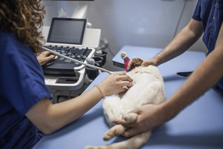
One of the biggest goals in veterinary medicine is to gather the information needed to make the best possible decisions while minimizing stress and risk for the patient.
When a pet is sick, it is natural for owners to want answers as quickly as possible. Whether a dog has been vomiting for several days, a cat has stopped eating, or routine blood work reveals unexpected abnormalities, the next step often involves determining exactly what is happening inside the body.
The challenge is that many illnesses produce similar symptoms.
A pet with pancreatitis may have symptoms that resemble liver disease. Intestinal inflammation may look similar to certain cancers. Gallbladder disease can mimic gastrointestinal disorders. Even heart disease may initially present as fatigue or decreased activity rather than obvious cardiac symptoms.
Because so many conditions overlap, veterinarians rely on advanced diagnostics to narrow the list of possibilities before recommending medications, surgery, or additional procedures.
This is where veterinary ultrasound and echocardiography have become invaluable.
Rather than immediately moving toward invasive testing, ultrasound often provides detailed information that helps veterinarians determine the safest, most appropriate path forward.
For many patients, advanced imaging answers important questions while avoiding unnecessary procedures.
Why Finding the Cause Matters More Than Treating the Symptoms
One of the most common misconceptions in veterinary medicine is that treating symptoms automatically solves the problem.
In reality, symptoms are only clues.
Vomiting is not a diagnosis.
Weight loss is not a diagnosis.
A heart murmur is not a diagnosis.
These findings simply indicate that something within the body deserves further investigation.
Treating vomiting without understanding its cause may temporarily improve comfort while allowing a more serious condition to continue progressing.
Likewise, prescribing medication for coughing without evaluating the heart may overlook cardiac disease that requires a completely different treatment approach.
Advanced imaging helps veterinarians identify the underlying problem rather than simply managing outward signs of illness.
This allows treatment plans to become far more targeted and effective.
Why Ultrasound Is Often Recommended Before Surgery
Many pet owners become understandably anxious when surgery is mentioned.
In some situations, surgery is absolutely necessary.
However, there are many cases where veterinarians first recommend ultrasound to determine whether an operation is actually needed.
For example, a dog experiencing repeated vomiting may have:
- Severe gastritis
- Pancreatitis
- An intestinal foreign body
- Intestinal inflammation
- Gallbladder disease
- A mass affecting the digestive tract
Several of these conditions may cause nearly identical symptoms.
Ultrasound allows veterinarians to evaluate the abdominal organs in real time and determine whether surgery is likely to benefit the patient or whether medical management is the better option.
This information helps avoid unnecessary procedures while ensuring pets that truly require surgery receive timely care.
Looking Beyond What X-Rays Can Show
Radiographs remain an important part of veterinary medicine.
They are excellent for evaluating bones, detecting fractures, assessing lung changes, and identifying certain abnormalities involving the chest or abdomen.
However, X-rays have limitations when it comes to soft tissues.
Two organs may appear similar on a radiograph despite having very different diseases.
Ultrasound provides additional detail by allowing veterinarians to examine:
- Organ structure
- Tissue texture
- Blood vessels
- Fluid accumulation
- Internal architecture
- Blood flow in selected structures
This deeper level of evaluation frequently changes the direction of a diagnostic workup.
Rather than simply identifying that an organ is enlarged, ultrasound helps explain why it is enlarged.
That distinction is often critical when deciding how to proceed.
Understanding When Biopsies Become Necessary
Another important role of ultrasound is helping veterinarians determine when tissue sampling may be appropriate.
Occasionally, imaging identifies an abnormal area that cannot be fully characterized through appearance alone.
Rather than performing exploratory surgery immediately, ultrasound may help guide minimally invasive procedures such as fine-needle aspiration.
These samples can often provide valuable information about:
- Inflammation
- Infection
- Benign growths
- Certain cancers
- Abnormal cell populations
In some cases, these procedures eliminate the need for more invasive surgery.
In others, they help surgeons plan procedures more effectively by identifying exactly where abnormalities are located.
The goal is always to gather the information needed while keeping the patient’s safety at the forefront.
How Ultrasound Helps Evaluate Multiple Organs at the Same Time
One advantage of veterinary ultrasound is that it rarely focuses on just one organ.
Although a pet may be referred because of elevated liver enzymes or chronic vomiting, the examination often includes a comprehensive evaluation of the abdomen.
This broader approach allows veterinarians to assess how multiple organ systems may be interacting.
For example, digestive symptoms may actually involve the pancreas, gallbladder, liver, intestines, or lymph nodes simultaneously.
Examining these structures together provides a much more complete understanding of the patient’s overall health.
This comprehensive evaluation is one reason ultrasound has become such an important diagnostic tool in both routine and complex medical cases.
Echocardiograms Provide Information That Physical Exams Cannot
Heart murmurs are commonly detected during routine wellness visits.
While listening to the heart with a stethoscope provides valuable information, it cannot reveal exactly how the heart is functioning.
An echocardiogram allows veterinarians to observe the heart while it is actively pumping blood.
This specialized ultrasound evaluates:
- Heart valves
- Chamber dimensions
- Blood flow
- Heart muscle thickness
- Pumping efficiency
- Structural abnormalities
For pets with suspected heart disease, this information helps determine whether changes are mild, progressive, or require immediate treatment.
It also helps veterinarians identify pets that can safely undergo anesthesia if another procedure becomes necessary.
Why Advanced Imaging Supports Better Long-Term Planning
Veterinary medicine is about more than diagnosing disease.
It is also about planning for the future.
Many chronic conditions require ongoing monitoring rather than one-time treatment.
Ultrasound and echocardiography allow veterinarians to compare studies over time, helping them evaluate whether disease remains stable or continues progressing.
This information supports decisions involving medication adjustments, follow-up testing, dietary management, and long-term care strategies.
For pets living with chronic illnesses, these regular assessments often become an important part of maintaining quality of life.
Every Diagnostic Decision Should Be Guided by the Individual Patient
No two patients are exactly alike.
Two dogs with similar symptoms may have completely different underlying diseases.
Two cats with identical blood work may require entirely different treatment plans.
Advanced imaging helps veterinarians move beyond assumptions by providing objective information about the patient’s internal health.
Rather than relying solely on symptoms, physical examinations, or laboratory values, ultrasound and echocardiography offer another layer of detail that often changes how medical decisions are made.
For many pets, that additional information is what transforms uncertainty into a clear diagnostic plan.
When Repeat Ultrasounds Become Part of Ongoing Care
Many pet owners think of ultrasound as a one-time diagnostic procedure that is only used to identify an illness.
In reality, advanced imaging often becomes an important part of monitoring a pet’s health over time.
Certain medical conditions require regular evaluation because they can change gradually, even when a pet appears stable at home.
Veterinarians may recommend repeat ultrasounds to monitor:
- Gallbladder disease
- Liver abnormalities
- Kidney disease
- Pancreatic conditions
- Bladder masses
- Splenic nodules
- Intestinal disease
- Benign cysts
- Certain types of tumors
Comparing current images with previous examinations allows veterinarians to detect subtle changes that may not yet be causing clinical signs.
A mass that has remained unchanged for twelve months may require a very different treatment plan than one that has doubled in size over the same period.
Likewise, a gallbladder that continues to function normally may simply require monitoring, while progressive changes could indicate the need for medical or surgical intervention.
Ultrasound provides a safe, non-invasive way to gather this information while minimizing stress for both pets and their owners.
Echocardiograms Help Guide More Than Heart Medications
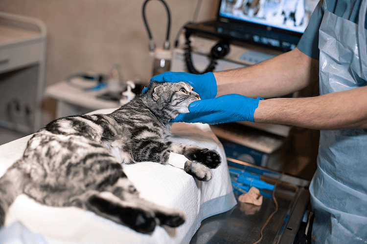
Many people associate echocardiograms solely with diagnosing heart disease.
While identifying cardiac disease is certainly one of their most important uses, echocardiography also helps veterinarians make decisions about many other aspects of patient care.
For example, if a pet requires anesthesia for dental treatment, surgery, or another procedure, understanding heart function beforehand may be essential.
An echocardiogram helps determine:
- Whether the heart is pumping efficiently
- Whether heart chambers are enlarged
- Whether valve disease is present
- Whether abnormal blood flow exists
- Whether anesthesia risks should be adjusted
This allows the veterinary team to create a treatment plan that is tailored to the individual patient’s cardiovascular health.
For pets with known heart disease, repeat echocardiograms also help determine when medications should be started, adjusted, or continued.
Rather than relying only on symptoms, veterinarians can evaluate measurable changes in heart function over time.
How Ultrasound Supports Collaborative Veterinary Care
Many veterinary patients receive care from more than one medical professional.
A primary care veterinarian may recommend additional imaging when a pet develops persistent symptoms or when specialized expertise is needed.
The ultrasound findings then become an important part of the patient’s overall medical record.
Diagnostic imaging can help support:
- Internal medicine consultations
- Surgical referrals
- Oncology treatment planning
- Cardiology evaluations
- Emergency medicine cases
- Chronic disease management
Because ultrasound findings can be shared with the referring veterinarian, every member of the medical team has access to detailed information that supports coordinated care.
This collaborative approach helps ensure that treatment recommendations are based on the most complete understanding of the patient’s condition.
What Pet Owners Can Expect During an Ultrasound Appointment
One of the most common concerns owners have is not knowing what happens during an ultrasound examination.
Fortunately, the procedure is generally straightforward and well tolerated by most dogs and cats.
Depending on the area being evaluated, a small amount of fur is usually clipped to allow better contact between the ultrasound probe and the skin.
A special gel is applied to improve image quality.
Most pets lie comfortably on a padded examination table while the sonographer systematically evaluates each organ.
Unlike many medical procedures, ultrasound is painless.
Many patients remain relaxed throughout the examination.
Some even become comfortable enough to rest during the scan.
Sedation is not routinely required, although it may occasionally be recommended for pets that are extremely anxious, painful, or unable to remain still.
The examination itself often takes between 30 and 60 minutes, depending on the complexity of the case and the structures being evaluated.
The Value of High-Quality Imaging Equipment
Veterinary imaging technology has advanced considerably over the past decade.
Modern ultrasound systems provide significantly higher image resolution than earlier generations of equipment.
Improved technology allows sonographers to visualize smaller anatomical structures and detect subtle abnormalities that previously may have been difficult to identify.
Some newer systems also provide enhanced Doppler imaging, which helps evaluate blood flow throughout organs and the heart.
While technology alone does not replace clinical expertise, it does allow experienced imaging professionals to gather more detailed diagnostic information.
Combining advanced equipment with skilled interpretation gives veterinarians greater confidence when developing treatment recommendations.
Ultrasound and Wellness Care Can Work Together
Many people associate ultrasound with sick pets, but advanced imaging also plays an important role in preventive healthcare.
For pets with breed-related health risks, chronic medical conditions, or previously identified abnormalities, ultrasound may become part of a proactive wellness strategy.
Rather than waiting until symptoms become severe, veterinarians may recommend imaging to monitor known conditions or investigate subtle changes detected during routine wellness examinations.
Examples include:
- Monitoring previously identified liver nodules
- Evaluating chronic kidney disease
- Following gallbladder abnormalities
- Monitoring heart murmurs
- Assessing changes identified on routine blood work
This proactive approach allows veterinarians to detect progression earlier and adjust treatment before complications develop.
Every Image Adds Another Piece to the Diagnostic Puzzle
Veterinary medicine rarely relies on a single diagnostic test.
Instead, veterinarians combine information from multiple sources to develop the clearest possible understanding of a patient’s health.
A physical examination provides valuable clinical observations.
Blood work evaluates organ function and overall health.
Radiographs help assess bones and certain internal structures.
Ultrasound and echocardiography provide another layer of information by allowing veterinarians to evaluate soft tissues, organ architecture, and heart function in real time.
Each diagnostic tool contributes a different perspective.
When combined, these findings help create a more complete picture that supports accurate diagnosis, individualized treatment plans, and long-term patient care.
Veterinary ultrasound and echocardiography are not simply imaging procedures.
They are valuable diagnostic resources that help veterinarians answer complex medical questions, monitor disease progression, and provide pets with care that is based on evidence rather than assumptions.