
Why Healthy Pets Sometimes Need Veterinary Ultrasounds and Echocardiograms: The Role of Advanced Imaging in Preventive Care
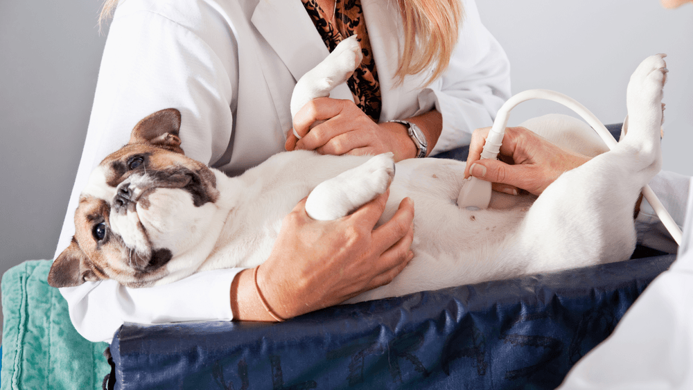
When most people think about veterinary ultrasounds or echocardiograms, they picture a pet that is obviously sick.
Perhaps a dog has been vomiting for several days, a cat has stopped eating, or routine blood work has uncovered unexpected abnormalities. In many of these situations, advanced imaging plays an essential role in helping veterinarians identify the cause of illness and develop an appropriate treatment plan.
However, diagnostic imaging is not only valuable after symptoms appear.
In modern veterinary medicine, preventive care has become increasingly focused on identifying health concerns before they progress into more serious conditions. Just as routine wellness examinations and laboratory testing help monitor your pet’s overall health, veterinary ultrasounds and echocardiograms can provide important information about internal organs and heart function—even when a pet appears healthy.
At Veterinary Diagnostics Center, we believe that advanced imaging is one of the most valuable tools available for both diagnosing disease and supporting lifelong wellness. By looking beyond what is visible during a physical examination, ultrasound and echocardiography help veterinarians make informed decisions that support earlier intervention, individualized care, and better long-term outcomes.
Preventive Care Means Looking Beyond Visible Symptoms
Dogs and cats have an incredible ability to compensate for illness.
Many pets continue eating, playing, and interacting with their families despite developing underlying medical conditions. By the time obvious symptoms become noticeable, disease may have been progressing silently for weeks, months, or even years.
This is especially true for conditions involving:
- Heart disease
- Liver disease
- Kidney disease
- Gallbladder disease
- Certain cancers
- Chronic intestinal disease
- Pancreatic disorders
Routine wellness visits are designed to detect these problems as early as possible. During these appointments, veterinarians evaluate your pet’s weight, body condition, heart and lung sounds, mobility, oral health, and overall physical condition.
Blood work provides another layer of valuable information by identifying changes in organ function that may not yet cause clinical signs.
Sometimes these tests raise questions that cannot be answered through laboratory results alone.
Advanced imaging helps bridge that gap.
Ultrasound Helps Reveal What Physical Exams Cannot
A veterinarian can often feel enlarged organs, detect abdominal discomfort, or hear changes in the heart during a physical examination.
What cannot be determined through touch or listening alone is exactly what those changes mean.
An abdominal ultrasound allows veterinarians to evaluate internal organs in remarkable detail, including:
- Liver
- Gallbladder
- Kidneys
- Spleen
- Pancreas
- Bladder
- Adrenal glands
- Intestinal tract
- Lymph nodes
Rather than simply identifying that an organ feels enlarged, ultrasound helps determine whether the underlying cause may involve inflammation, cysts, fluid accumulation, structural abnormalities, or masses.
This level of detail supports more accurate diagnoses while helping veterinarians choose the most appropriate next steps.
Baseline Imaging Creates a Valuable Point of Comparison
One benefit of preventive imaging that many pet owners have never considered is the value of establishing a baseline.
Every pet is unique.
Minor anatomical variations may be completely normal for one individual while representing significant change in another.
Having baseline ultrasound or echocardiogram findings available can become extremely valuable if medical concerns arise later.
Future examinations can be compared with earlier studies to determine:
- Whether an organ has changed in size
- Whether a previously identified nodule has grown
- Whether gallbladder abnormalities are progressing
- Whether kidney disease remains stable
- Whether heart structure has changed over time
Instead of relying on a single snapshot, veterinarians can evaluate trends.
Those trends often provide better insight into a pet’s overall health than one examination alone.
Breed-Related Health Risks Can Influence Imaging Recommendations
Some dogs and cats have inherited predispositions that increase their risk for developing specific medical conditions.
While every pet is an individual, breed history can help veterinarians determine whether additional monitoring may be beneficial.
For example, certain breeds are more likely to develop heart disease, while others are predisposed to gallbladder disease, liver abnormalities, or inherited kidney conditions.
In these situations, advanced imaging may be recommended before obvious symptoms develop.
The goal is not to assume disease is present.
Instead, it is to establish a clearer understanding of the patient’s health so any future changes can be recognized more quickly.
Preventive imaging becomes another tool that supports personalized veterinary care.
Echocardiograms Can Detect Changes Before Heart Disease Becomes Advanced
Heart murmurs are one of the most common reasons veterinarians recommend an echocardiogram.
However, cardiac imaging may also be valuable for pets with breed-related risk factors, subtle changes in stamina, or abnormal findings during routine wellness examinations.
An echocardiogram allows veterinarians to evaluate:
- Heart valve function
- Chamber dimensions
- Heart muscle thickness
- Blood flow patterns
- Pumping efficiency
- Structural abnormalities
These measurements help determine whether the heart is functioning normally or whether early changes deserve continued monitoring.
In some cases, identifying cardiac disease before symptoms appear allows veterinarians to recommend lifestyle adjustments, periodic rechecks, or medical management earlier in the disease process.
Earlier detection often provides more opportunities to support long-term heart health.
Wellness Imaging Is About More Than Finding Disease
Preventive ultrasound is not performed because veterinarians expect every patient to have a serious illness.
In many cases, the examination confirms that internal organs appear healthy and functioning normally.
That reassurance is valuable.
Knowing that imaging findings are normal gives both veterinarians and pet owners greater confidence when interpreting future laboratory results or monitoring changes over time.
When abnormalities are identified, they are often detected at a stage when additional treatment options remain available.
This proactive approach reflects one of the most important goals of modern veterinary medicine: helping pets stay healthier for as long as possible rather than simply responding after disease has become advanced.
Preventive Imaging Becomes Even More Valuable as Pets Age
As dogs and cats enter their senior years, routine wellness care often becomes more comprehensive.
While many older pets continue to enjoy active, happy lives, aging naturally increases the likelihood of developing chronic medical conditions. Some changes occur gradually, making them difficult to detect during the early stages.
Preventive ultrasound and echocardiography allow veterinarians to monitor aging pets more closely without waiting for significant symptoms to develop.
Senior wellness imaging may be recommended to evaluate:
- Changes identified during routine blood work
- Newly detected heart murmurs
- Mild weight loss
- Reduced energy levels
- Increased thirst or urination
- Changes in appetite
- Previously diagnosed chronic diseases
By identifying subtle structural changes early, veterinarians can often recommend monitoring plans or treatment strategies before more advanced disease affects a pet’s quality of life.
Monitoring Existing Conditions With Confidence
Not every ultrasound is performed to diagnose a new illness.
Many examinations are scheduled because veterinarians want to understand whether a known condition is remaining stable.
For example, a pet diagnosed with chronic kidney disease may benefit from periodic imaging to monitor kidney size, blood flow, and structural changes.
Likewise, pets with liver disease, gallbladder abnormalities, pancreatic disorders, or previously identified nodules may undergo repeat ultrasounds to compare findings over time.
Rather than relying solely on clinical signs, veterinarians can evaluate objective changes that help determine whether current treatment remains appropriate.
This approach supports individualized medical care while reducing unnecessary guesswork.
Wellness Blood Work and Imaging Work Best Together
Routine laboratory testing is an essential part of preventive medicine, but blood work and ultrasound answer different questions.
Blood tests measure how organs are functioning.
Ultrasound evaluates how those organs appear structurally.
For example, elevated liver enzymes may indicate inflammation or stress affecting the liver.
Ultrasound helps determine whether those changes are associated with liver enlargement, gallbladder disease, masses, cysts, or another structural abnormality.
Similarly, changes in kidney values may prompt imaging that identifies chronic degeneration, kidney stones, congenital abnormalities, or urinary tract disease.
When interpreted together, laboratory testing and ultrasound provide a more complete understanding of a patient’s overall health.
Echocardiograms Help Veterinarians Make Safer Medical Decisions
Heart health influences many aspects of veterinary medicine.
An echocardiogram is not only valuable for diagnosing heart disease—it also helps veterinarians make informed decisions before procedures that require anesthesia.
Understanding cardiac function before surgery, dental treatment, or other interventions allows the veterinary team to develop an anesthetic plan tailored to the individual patient.
An echocardiogram can provide information about:
- Valve function
- Chamber enlargement
- Blood flow through the heart
- Pumping efficiency
- Heart muscle thickness
- Congenital abnormalities
These findings help determine whether additional precautions or monitoring are appropriate during treatment.
Why Experience Matters in Veterinary Ultrasound
Ultrasound is one of the most operator-dependent diagnostic tools used in veterinary medicine.
Obtaining high-quality images requires advanced equipment, but interpreting those images requires experience, training, and attention to detail.
Every scan is dynamic.
The sonographer evaluates each organ from multiple angles while assessing tissue appearance, blood flow, organ relationships, and subtle abnormalities that could influence diagnosis.
Even findings that appear minor may become clinically important when considered alongside laboratory results and a pet’s medical history.
At Veterinary Diagnostics Center, advanced imaging is performed with a commitment to accuracy, collaboration, and thorough evaluation, providing referring veterinarians with meaningful diagnostic information to support patient care.
Supporting Collaborative Veterinary Care
Advanced imaging is often part of a larger healthcare team.
Primary care veterinarians, emergency clinicians, internal medicine specialists, surgeons, and cardiologists each contribute valuable expertise when managing complex cases.
Ultrasound and echocardiography provide detailed information that can be shared with referring veterinarians, ensuring everyone involved has access to the same diagnostic findings.
This collaborative approach helps guide decisions involving:
- Medical management
- Surgical planning
- Specialist referrals
- Long-term monitoring
- Follow-up imaging
- Ongoing wellness care
Working together allows veterinary teams to create individualized treatment plans based on a comprehensive understanding of each patient’s condition.
Advanced Imaging Supports Lifelong Wellness
Preventive medicine is about more than preventing disease.
It is about understanding each patient’s health well enough to recognize meaningful change when it occurs.
Veterinary ultrasounds and echocardiograms provide information that cannot be obtained through physical examination alone.
Whether confirming normal organ health, monitoring a chronic condition, evaluating heart function, or investigating subtle laboratory abnormalities, advanced imaging helps veterinarians make evidence-based decisions with confidence.
For many pets, these examinations offer reassurance that everything appears healthy.
For others, they provide the opportunity to identify disease earlier, monitor progression more accurately, and begin treatment before serious complications develop.
Every image contributes another piece to the larger picture of your pet’s health, supporting thoughtful medical care throughout every stage of life.