Why Some Health Problems in Pets Go Undetected Until Advanced Imaging Reveals Them
One of the most frustrating situations in veterinary medicine occurs when a pet is clearly not acting like themselves, yet initial testing does not provide a clear answer.
The dog that suddenly seems tired.
The cat that gradually loses weight.
The pet that continues vomiting despite treatment.
The senior dog whose blood work looks only mildly abnormal but somehow still seems unwell.
These situations happen every day in veterinary practices across the country.
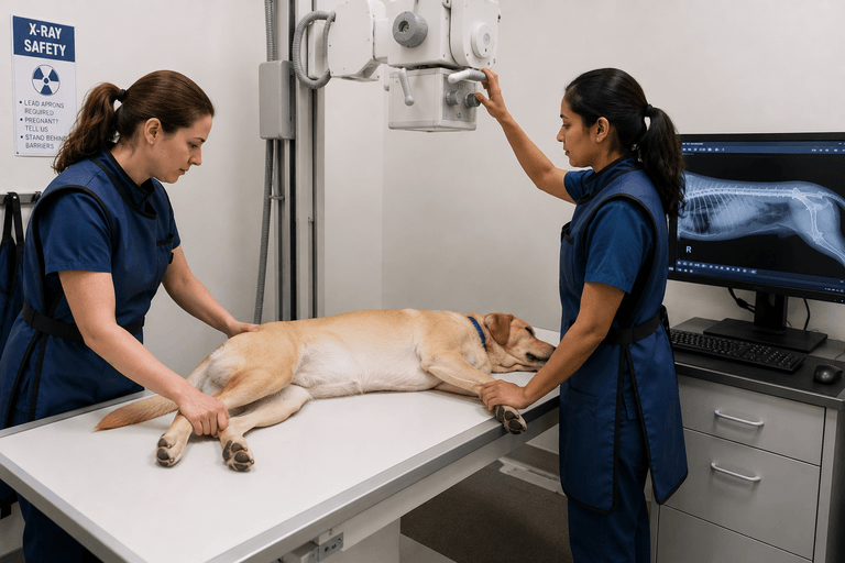
While physical examinations, blood tests, and X-rays provide valuable information, they do not always tell the entire story. Sometimes the answers are hidden deeper within the body, requiring more advanced imaging to uncover what is truly happening.
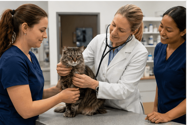
This is where veterinary ultrasound and echocardiography often become some of the most important diagnostic tools available.
Advanced imaging allows veterinarians to move beyond symptoms and begin evaluating the organs, tissues, and structures that may be contributing to a pet’s condition.
In many cases, ultrasound reveals problems that would otherwise remain undetected until they become far more serious.
Why Symptoms Alone Do Not Always Tell the Full Story
Pets are remarkably skilled at hiding illness.
Many dogs and cats naturally compensate for disease long before obvious symptoms appear. By the time owners notice a problem, the condition may have been developing for weeks or even months.
A pet experiencing liver disease may simply seem less energetic.
A dog with developing heart disease may only show subtle exercise intolerance.
A cat with intestinal disease may occasionally vomit but otherwise appear normal.
Because these signs are often vague, veterinarians rely on diagnostics to uncover what cannot be seen externally.
The challenge is that even standard diagnostics sometimes have limitations.
Blood work may indicate that something is wrong without identifying exactly where the problem exists.
X-rays may reveal changes in organ size but not what is happening inside those organs.
This is often the point where advanced imaging becomes the next logical step.
How Veterinary Ultrasound Provides a Different Level of Information
One of the greatest advantages of veterinary ultrasound is its ability to evaluate soft tissue structures in real time.
Rather than simply viewing an organ’s outline, veterinarians can assess its internal appearance, movement, texture, and surrounding tissues.
Ultrasound is commonly used to evaluate:
- Liver
- Gallbladder
- Kidneys
- Pancreas
- Spleen
- Bladder
- Intestinal tract
- Lymph nodes
- Adrenal glands
This additional detail often helps veterinarians answer questions that other diagnostic methods cannot.
For example, an X-ray may show an enlarged liver.
An ultrasound can help determine whether that enlargement is associated with inflammation, nodules, gallbladder disease, masses, or other abnormalities.
The difference can dramatically affect treatment recommendations.
Conditions That Frequently Benefit From Ultrasound Evaluation
Persistent Digestive Problems
Chronic vomiting, diarrhea, decreased appetite, and unexplained weight loss are among the most common reasons veterinarians recommend abdominal ultrasound.
Many digestive disorders create similar symptoms, making it difficult to identify the underlying cause through observation alone.
Ultrasound may help identify:
- Intestinal thickening
- Foreign material
- Gallbladder abnormalities
- Pancreatic disease
- Enlarged lymph nodes
- Masses
- Fluid accumulation
In some cases, multiple organ systems may be contributing to the same symptoms.
Without advanced imaging, those connections can be difficult to recognize.
Abnormal Blood Work Without Obvious Symptoms
One of the most surprising situations for pet owners occurs when blood work reveals abnormalities even though their pet appears relatively normal.
Elevated liver values are a common example.
Many owners assume this automatically means liver disease.
However, ultrasound frequently reveals that the gallbladder, pancreas, intestines, or another organ may actually be contributing to the abnormal laboratory results.
This is why imaging often becomes a critical piece of the diagnostic puzzle.
Urinary Tract Disorders
Urinary issues can quickly become serious, especially when obstruction is involved.
Ultrasound can help identify:
- Bladder stones
- Kidney abnormalities
- Urinary tract obstruction
- Bladder masses
- Chronic inflammation
In emergency situations, rapid identification of these conditions can significantly influence treatment decisions and outcomes.
The Unique Role of Echocardiograms in Veterinary Medicine
While abdominal ultrasound evaluates internal organs, an echocardiogram focuses specifically on the heart.
An echocardiogram is essentially an ultrasound of the heart that allows veterinarians to evaluate cardiac function in real time.
This imaging provides information that cannot be obtained through physical examination alone.
Veterinarians can assess:
- Heart chamber size
- Valve function
- Blood flow patterns
- Heart muscle thickness
- Pumping efficiency
- Pulmonary pressures
For patients with suspected heart disease, echocardiography remains the gold standard for evaluating cardiac structure and performance.
Signs That May Indicate an Echocardiogram Is Needed
Many pets are referred for echocardiography after a veterinarian detects a heart murmur or arrhythmia during a routine examination.
Additional reasons may include:
- Coughing
- Difficulty breathing
- Exercise intolerance
- Collapse episodes
- Weakness
- Changes in stamina
These symptoms do not always mean a pet has heart disease.
However, they often warrant a closer look at how the heart is functioning.
Early detection allows veterinarians to monitor disease progression and develop treatment plans before severe complications occur.
Why Experience Matters in Veterinary Imaging
Ultrasound is unlike many other imaging technologies.
The quality of the examination depends heavily on the experience of the individual performing the scan.
A skilled sonographer does more than simply capture images.
They actively evaluate structures in real time, investigate abnormalities, and adjust their approach based on what they discover during the examination.
Experienced imaging professionals understand:
- Which areas require additional evaluation
- How disease patterns typically appear
- How to recognize subtle abnormalities
- When findings warrant further investigation
This level of expertise can make a significant difference in diagnostic accuracy.
When Imaging Reveals Unexpected Findings
One of the most valuable aspects of advanced imaging is its ability to uncover conditions that were not originally suspected.
Veterinarians frequently discover findings unrelated to the initial reason for referral.
Examples may include:
- Splenic masses
- Gallbladder disease
- Bladder stones
- Enlarged lymph nodes
- Early heart disease
- Organ abnormalities
Sometimes these incidental discoveries allow treatment to begin before the condition progresses into a more serious problem.
Early detection remains one of the most powerful advantages of modern veterinary diagnostics.
The Goal Is Better Answers, Not Just More Testing
Pet owners sometimes worry that additional diagnostics simply create more questions.
The reality is that advanced imaging often helps veterinarians eliminate uncertainty.
Even when ultrasound does not provide a definitive diagnosis, it frequently narrows the list of possibilities and helps guide the next steps.
That information can prevent unnecessary treatments, improve medical decision-making, and provide clearer recommendations for both veterinarians and pet owners.
Ultimately, the goal is not simply to perform more tests.
The goal is to understand what is happening inside the patient as accurately as possible.
And in many cases, veterinary ultrasound and echocardiography provide the clarity needed to move forward with confidence.