
When an Ultrasound Finds an Abnormality: How Imaging Can Guide the Next Step in Your Pet’s Diagnosis
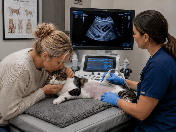
When a veterinarian recommends a veterinary ultrasound, pet owners understandably hope the scan will provide an immediate answer.
Sometimes it does.
Other times, the ultrasound reveals something that requires a closer look.
A mass may be visible. An organ may have an unusual appearance. A lymph node may look different from what is expected. There may be an area of inflammation or another structural change that cannot be fully explained by the pet’s symptoms or blood work alone.
Finding an abnormality is an important step, but it may not be the final step.
In these situations, veterinary ultrasound can do more than show veterinarians that something is different. The detailed, real-time images can help the veterinary team understand where the abnormality is located, what structures surround it, and whether additional diagnostic testing may be appropriate.
This is one of the ways advanced veterinary imaging becomes part of a larger diagnostic process.
At Veterinary Diagnostics Centers, our approach to ultrasound is centered on providing veterinarians with meaningful information that can help guide the next medical decision. When an abnormality requires additional investigation, imaging can help provide a clearer roadmap for what comes next.
An Abnormal Ultrasound Finding Is Not Always a Diagnosis
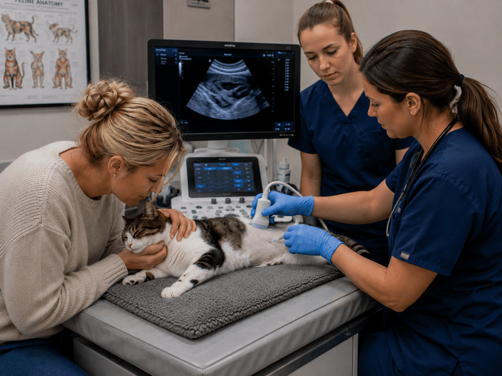
One of the most important things for pet owners to understand is that seeing an abnormality on an ultrasound does not always tell us exactly what that abnormality is.
Ultrasound can reveal a mass, abnormal lymph nodes, intestinal changes, gallbladder abnormalities, fluid, or other structural findings. However, imaging alone may not determine the exact type of disease or whether a particular mass is cancerous.
That distinction matters.
For example, a veterinarian may identify a mass during an abdominal ultrasound. The image can provide important information about the mass’s location and appearance, but determining its exact cellular characteristics may require another diagnostic method.
Depending on the findings and the patient’s individual situation, the veterinarian may recommend additional testing such as:
- Fine-needle aspiration
- Cytology
- Biopsy
- Additional blood work
- Follow-up imaging
- CT or another advanced diagnostic test
The ultrasound therefore becomes an important part of the diagnostic pathway rather than being viewed as an isolated test.
Why Location Matters When an Abnormality Is Found
Knowing that something abnormal is present is helpful.
Knowing where it is can be even more useful.
The abdomen contains many organs and structures that are located close together. A mass or abnormal area may originate from one organ while appearing close to another. Understanding its location and relationship to surrounding structures can help veterinarians determine how best to investigate it.
Veterinary ultrasound provides real-time views of organs and tissues, including the liver, gallbladder, kidneys, bladder, pancreas, spleen, intestinal tract, adrenal glands, and lymph nodes. This allows the imaging examination to provide more context around an abnormal finding.
Instead of simply documenting that an abnormality exists, the veterinary team can evaluate its relationship to the surrounding anatomy.
That information can become particularly valuable when additional diagnostics are being considered.
Ultrasound Can Help Identify the Best Area to Investigate
When additional sampling is appropriate, the goal is to obtain useful diagnostic material while minimizing unnecessary disruption to the patient.
Ultrasound can help visualize the area being investigated in real time.
This is important because internal structures are not always exactly where a veterinarian might expect them to be based solely on an external examination or a two-dimensional image.
During an ultrasound examination, the sonographer can observe the relevant anatomy while the imaging is being performed.
This provides the veterinary team with a more detailed understanding of the area being evaluated.
When a fine-needle aspiration or another sampling procedure is recommended, that information can help guide the diagnostic approach.
What Is a Fine-Needle Aspiration?
A fine-needle aspiration, often called an FNA, involves using a small needle to collect cells from an abnormal area.
Those cells can then be evaluated through cytology.
The purpose is to obtain additional information about what is happening at the cellular level.
This can be particularly useful when an ultrasound identifies a mass, enlarged lymph node, or another abnormal structure that cannot be fully characterized through imaging alone.
Ultrasound does not replace cytology.
Instead, the two can work together.
The ultrasound identifies and characterizes the area of concern, while cytology may provide additional information about the cells within that area.
The source material specifically lists fine-needle aspiration and cytology among the additional diagnostics that may be recommended after an ultrasound identifies an abnormality.
Why Imaging and Sampling Can Complement Each Other
Think of diagnostic imaging as looking at the bigger picture.
An ultrasound can show:
- Where an abnormality is located
- Which organ or structure is involved
- The appearance of surrounding tissues
- Whether other abnormalities are visible
- Whether fluid or other changes are present
Sampling can provide a different type of information.
It can help veterinarians investigate what is happening at the cellular level.
Neither type of information necessarily replaces the other.
Instead, combining them can give the veterinary team a more complete understanding of a patient’s condition.
This is especially important because some diseases can look similar on imaging.
Two masses may have a similar appearance while having very different underlying causes.
That is why a veterinarian may recommend additional testing even after an ultrasound has clearly identified an abnormality.
The Goal Is Not Always to Sample Everything
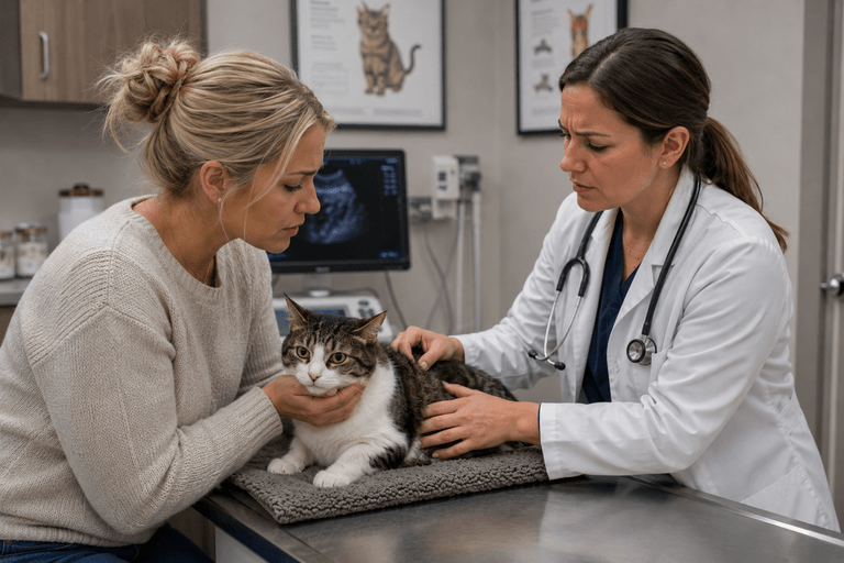
Finding an abnormality does not automatically mean that every patient needs a biopsy or needle sample.
The decision depends on the individual case.
Veterinarians consider factors such as:
- The patient’s symptoms
- The location of the abnormality
- The appearance of the abnormality
- The pet’s overall health
- Blood work
- Previous diagnostic results
- Whether the finding appears stable or changing
- Whether the result would change treatment
This is another reason veterinary imaging should be interpreted within the context of the entire patient.
An ultrasound finding does not exist in isolation.
The veterinary team considers the imaging alongside the pet’s medical history and other diagnostic information.
Ultrasound Can Help Distinguish Between Different Diagnostic Possibilities
Some clinical signs can have many possible causes.
A pet may lose weight because of gastrointestinal disease, organ dysfunction, inflammation, or another underlying condition.
Similarly, an abnormal laboratory result may indicate a problem without showing where the problem is located.
Ultrasound provides an opportunity to investigate internal structures directly.
The source material explains that ultrasound can help veterinarians determine whether disease appears localized or widespread, which organs are involved, and whether additional testing is necessary.
This information can help narrow the diagnostic pathway.
Rather than approaching every possibility equally, veterinarians may be able to focus attention on the areas that imaging identifies as most relevant.
A More Detailed Look Can Change the Next Decision
One of the advantages of ultrasound is that the examination happens in real time.
The sonographer isn’t limited to one predetermined image.
If an area looks unusual, additional views can be obtained.
If an organ appears different from what was expected, it can be examined more closely.
If another abnormality becomes visible, the examination can be adjusted accordingly.
The existing transcript explains that ultrasound requires real-time technical decision-making and that the person performing the examination may need to adjust the scan based on what is seen.
This becomes especially important when the next diagnostic step depends on understanding exactly what is happening in a particular area.
Ultrasound Can Help With More Than Masses
While masses are an important example, ultrasound-guided diagnostic evaluation can be relevant to many types of abnormalities.
Depending on the patient’s condition, veterinarians may investigate:
- Enlarged lymph nodes
- Abnormal organs
- Fluid collections
- Focal lesions
- Certain inflammatory changes
- Abnormal tissue structures
- Other areas that require additional evaluation
The objective is always to gather useful information while considering the patient’s individual needs.
An abnormal finding does not automatically mean something severe is present.
But it does give the veterinary team another piece of information to consider.
What Happens After a Sample Is Collected?
If additional sampling is performed, the material collected is typically submitted for further evaluation.
Cytology examines cells obtained from the sample.
A biopsy involves obtaining tissue for more detailed examination.
The appropriate test depends on what the veterinarian is trying to determine.
This distinction is important because pet owners sometimes expect one test to answer every question.
Veterinary medicine often works in stages.
One diagnostic test may identify an area of concern.
A second test may characterize it.
A third may help determine the most appropriate treatment.
The process may look longer than simply receiving one final answer, but each step is intended to add useful information.
Ultrasound Can Help Veterinarians Make More Informed Decisions About Surgery
Sometimes an abnormality identified during ultrasound leads to surgery.
In other situations, imaging may provide information that helps determine that surgery is not immediately necessary.
The source material explains that ultrasound can help veterinarians assess whether surgery is needed, whether disease appears localized or widespread, which organs are involved, and whether additional testing is necessary.
This can be valuable when deciding how aggressively to approach a condition.
For example, understanding the location and extent of an abnormality may help a veterinarian determine what additional information is needed before recommending a procedure.
The objective is not to avoid surgery at all costs.
The objective is to make sure that when surgery is recommended, the veterinary team has as much relevant information as possible.
Better Diagnostic Information Can Improve Treatment Planning
A diagnosis is valuable because it influences what happens next.
Different diseases can require very different treatments.
Medical management may be appropriate for one condition.
Surgery may be appropriate for another.
Monitoring may make sense in a third situation.
Additional testing may be necessary before any treatment decision can be made.
This is why the information gathered through veterinary ultrasound can have implications beyond the imaging appointment itself.
The scan may help shape the next stage of the patient’s care.
It can provide veterinarians with information that supports more targeted decisions rather than relying solely on symptoms or assumptions.
Why the Imaging Process Matters
High-quality ultrasound depends on more than simply having an ultrasound machine.
Ultrasound is highly operator-dependent because the examination involves hands-on scanning and real-time decision-making. The source material emphasizes that an experienced sonographer needs to recognize subtle abnormalities, adjust imaging angles, track difficult structures, and focus on areas that may be important based on the patient’s symptoms.
This is particularly relevant when an abnormality may require additional investigation.
The imaging needs to provide enough information for the veterinary team to understand the area being evaluated.
The person performing the scan plays an important role in obtaining that information.
The Patient’s Entire Medical History Still Matters
Even the most detailed ultrasound does not replace the rest of a pet’s medical history.
Before an examination, the imaging team may review:
- Symptoms
- Previous diagnostics
- Blood work
- X-rays
- Medical history
The source material explains that this information helps guide the ultrasound examination.
This context allows the imaging findings to be interpreted more appropriately.
For example, an abnormality discovered during a scan may have greater significance when it corresponds with the patient’s symptoms or laboratory results.
Alternatively, an imaging finding may be unrelated to the original concern.
Understanding the entire clinical picture helps veterinarians determine what deserves additional attention.
What If the Ultrasound Does Not Find the Cause?
Not every diagnostic ultrasound produces a clear explanation.
That does not mean the examination was unsuccessful.
Ultrasound may help rule out major concerns, narrow down possibilities, or show that certain organs appear normal.
This information can still be useful.
The source material notes that ultrasound may confirm disease, rule out major concerns, detect masses or tumors, identify inflammation, find fluid or internal bleeding, and locate obstructions—but it cannot always determine the exact type or microscopic severity of a disease.
When the cause remains unclear, the veterinary team can use the available information to determine what should be investigated next.
Sometimes that means another diagnostic test.
Sometimes it means monitoring.
Sometimes it means returning to the original clinical question and considering another possibility.
Advanced Imaging Is About Building a Better Diagnostic Picture
A veterinary ultrasound does not need to answer every question to be valuable.
Sometimes the most important contribution is identifying an abnormality that would otherwise have remained hidden.
Sometimes it helps locate the source of a problem.
Sometimes it provides information that makes additional testing more focused.
And sometimes it helps veterinarians determine that an area of concern does not require immediate intervention.
This is the larger role of advanced veterinary imaging.
At Veterinary Diagnostics Centers, ultrasounds and echocardiograms are part of a broader diagnostic process designed to help veterinarians understand what is happening inside their patients and make informed decisions about the care that follows.
When an ultrasound reveals something unexpected, the next step is not necessarily a reason for alarm.
It is an opportunity to gather more information.
And in veterinary medicine, having the right information can make all the difference in determining what a pet needs next.