
Why Every Veterinary Ultrasound Is Different: How Targeted Imaging Helps Answer the Right Questions
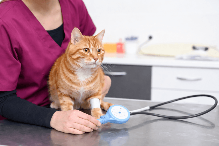
When a veterinarian recommends an ultrasound, many pet owners imagine a fairly straightforward process: a probe is placed on the abdomen, images are taken, and the results reveal what is wrong.
In reality, veterinary ultrasound is much more thoughtful and individualized than that.
The reason a dog or cat is being scanned matters. The patient’s medical history matters. Previous blood work, radiographs, physical examination findings, and even the veterinarian’s specific concerns can influence how the examination is performed.
At Veterinary Diagnostics Centers, we understand that an ultrasound is not simply about collecting a set of pictures. It is about asking the right diagnostic questions and using real-time imaging to investigate those questions as thoroughly as possible.
That distinction is important because every patient presents a different medical puzzle.
A pet with abnormal liver values may need a different diagnostic focus than a pet with chronic gastrointestinal symptoms. A patient being evaluated for an abdominal mass may require a different approach than a pet being assessed for urinary disease. And an echocardiogram is not simply an abdominal ultrasound performed in a different location—it is a specialized cardiac examination designed to evaluate the structure and function of the heart.
Understanding this individualized approach can help pet owners better appreciate what advanced veterinary imaging can contribute to their pet’s care.
Veterinary Ultrasound Is More Than Taking Pictures
Ultrasound is a real-time imaging technique.
Unlike an image that is captured once and reviewed later, the sonographer is actively observing structures while the examination is happening. The person performing the scan can change the angle, adjust the view, follow an organ, compare structures, and investigate something that looks different from what was initially expected.
That real-time aspect is one of the reasons veterinary ultrasound can provide such valuable information.
The examination is guided by the patient’s clinical situation.
For example, if a veterinarian is concerned about gastrointestinal disease, the scan may involve particularly careful evaluation of the stomach and intestinal tract. If urinary disease is suspected, the kidneys and bladder become important areas of attention. If abnormal liver values are present, the liver and gallbladder may receive particular consideration.
The goal is not simply to move a probe from one location to another.
The goal is to understand what is happening inside the patient.
The Clinical Question Helps Shape the Examination
A good diagnostic examination begins before the ultrasound machine is turned on.
Veterinary teams may review:
● The reason for the referral
● Current symptoms
● Duration of the problem
● Previous blood work
● Radiographs
● Medical history
● Previous imaging
● Known medical conditions
This information provides context.
The same ultrasound finding can have different significance depending on why the patient was referred.
A small abnormality in one patient may be incidental, while a similar finding in another patient may be directly relevant to the clinical concern.
This is why diagnostic imaging works best when the imaging team understands the patient rather than simply reviewing images in isolation.
The transcript supporting this article emphasizes that strong diagnostic work requires knowing what you are looking for in a particular case and being able to react in real time during the scan.
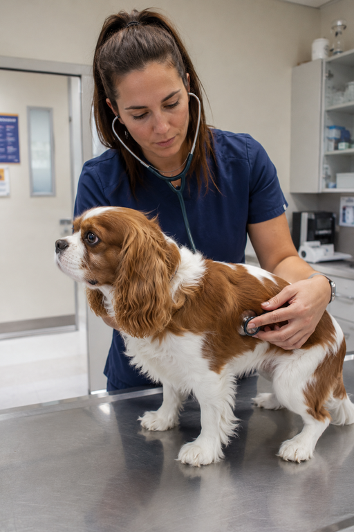
One Abdomen Can Tell Many Different Stories
The abdomen contains multiple organs that work together, and disease affecting one system can sometimes create symptoms that appear to originate somewhere else.
This makes abdominal ultrasound particularly useful for investigating complicated cases.
A veterinary ultrasound can evaluate structures including the:
● Liver
● Gallbladder
● Kidneys
● Spleen
● Pancreas
● Urinary bladder
● Stomach
● Small intestines
● Large intestines
● Adrenal glands
● Lymph nodes
These organs can be assessed for changes in size, shape, architecture, and other abnormalities.
The examination can therefore provide information that helps veterinarians understand whether a problem appears localized to one organ or involves multiple structures.
This is especially valuable when the original clinical signs are vague.
A pet may simply seem tired.
Another may have decreased appetite.
A third may have abnormal laboratory results without obvious symptoms at home. The imaging process helps turn those broad concerns into more specific questions.
When the First Finding Changes the Examination
One of the most important characteristics of ultrasound is that the examination can evolve as new information appears.
Imagine that a veterinarian is initially concerned about one organ system. During the scan, another abnormality is identified.
That finding may change what the sonographer needs to examine next.
Instead of following a rigid sequence without deviation, the examination can adapt.
This is one reason ultrasound is considered highly operator-dependent. The person performing the scan must recognize meaningful abnormalities and know when additional attention is warranted. Veterinary ultrasound therefore requires more than technical familiarity with the equipment. It requires clinical awareness and the ability to make decisions while the examination is taking place.
This flexibility is especially valuable in complicated medical cases where the original concern does not necessarily explain everything happening inside the patient.
Ultrasound Can Help Connect Findings That Seem Unrelated
Sometimes a pet’s symptoms appear disconnected.
For example, a patient may have poor appetite and abnormal liver values. Another may have weight loss and changes in gastrointestinal function. A third may have urinary symptoms alongside changes in kidney-related laboratory values.
An ultrasound can help veterinarians look at the relevant organ systems together.
Instead of evaluating each symptom as an isolated problem, imaging can provide a broader view of the internal structures involved.
This does not mean ultrasound always provides one definitive answer.
The source material makes an important distinction here: ultrasound may identify where a problem is located or reveal an abnormality that requires further investigation, but additional diagnostics may still be necessary.
That is not a weakness of ultrasound.
It is part of responsible diagnostic medicine.
A Finding Does Not Always Mean the Same Thing as a Diagnosis
One of the most important expectations for pet owners is understanding the difference between an imaging finding and a final diagnosis.
An ultrasound may identify a mass.
That does not automatically determine whether the mass is benign or malignant. It may show changes involving an organ.
That does not necessarily identify the exact microscopic disease responsible. Additional testing may be recommended depending on what the imaging reveals.
These next steps can include:
● Fine-needle aspiration
● Cytology
● Biopsy
● Additional laboratory testing
● Repeat imaging
● Other advanced diagnostics
The transcript specifically addresses this point: sometimes ultrasound answers an important question by identifying where the problem is, while further testing is needed to determine exactly what the disease is.
For pet owners, this distinction can make the diagnostic process easier to understand. The purpose of imaging is not always to produce one final answer immediately. Sometimes the most valuable result is knowing what needs to be investigated next.
Why Echocardiograms Require a Different Kind of Focus
An echocardiogram is a cardiac ultrasound, but it should not be thought of simply as an abdominal ultrasound focused on the heart.
The heart requires its own specialized examination because veterinarians are evaluating movement, blood flow, valves, chambers, wall thicknesses, and cardiac function.
The transcript describes echocardiography as a way to evaluate the heart’s valves, blood flow, velocities, directions, and wall thicknesses.
During a cardiac examination, multiple views and measurements may be obtained rather than relying on one still image.
This provides a much more complete assessment of cardiac health.
For example, an echocardiogram can help veterinarians evaluate:
● Heart chamber dimensions
● Valve function
● Heart wall thickness
● Blood flow
● Cardiac muscle
● Pumping function
● Certain pressure measurements
● Structural abnormalities
This level of detail is one reason echocardiography is so valuable when heart disease is suspected.
An Echocardiogram Can Answer Questions a Chest X-Ray Cannot
Chest radiographs remain an important diagnostic tool, but they provide different information from echocardiography.
A chest X-ray can show the overall size and shape of the heart and provide information about the lungs and chest.
An echocardiogram allows veterinarians to examine the heart itself in much greater detail.
The source material specifically explains that radiographs show the general size and shape of the heart, while echocardiography allows evaluation of internal structures such as valves and wall thicknesses.
This difference matters because heart disease is not defined solely by how large the heart looks.
A heart may have structural or functional abnormalities that require a closer evaluation.
Why the Same Heart Murmur Can Lead to Different Recommendations
A heart murmur is a finding, not a complete diagnosis.
When a veterinarian hears a murmur, the next question is what is causing it and whether it has clinical significance.
An echocardiogram can provide information about the heart’s structure and function that helps
answer that question.
The examination may help determine:
● Whether structural heart disease is present
● Which cardiac structures are affected
● How significant the changes appear
● Whether monitoring is appropriate
● Whether treatment may be recommended
The source material notes that echocardiography can be particularly important when a murmur or arrhythmia is identified and when anesthesia safety needs to be considered.
This allows care to be based on what is actually happening inside the heart rather than on the sound of the murmur alone.
Advanced Imaging Works Best as Part of a Larger Diagnostic Picture
Veterinary ultrasound and echocardiography are powerful tools, but they are not intended to replace every other diagnostic method.
The most useful diagnostic process combines information.
A veterinarian may begin with a physical examination and basic laboratory testing. Depending on the findings, radiographs may provide additional information. Ultrasound or echocardiography can then answer questions that those earlier diagnostics cannot fully address.
The transcript emphasizes that advanced diagnostics should fit into the broader diagnostic process rather than automatically being the first step for every case.
That approach is important for both medical accuracy and responsible patient care. The goal is not to perform every possible test.
The goal is to perform the right test at the right time to answer the most important clinical question.
What Experience Adds to the Examination
Because the examination happens in real time, experience can influence what gets investigated, which structures receive additional attention, and how unexpected findings are
followed.
High-quality ultrasound requires both appropriate technology and skilled interpretation.
The transcript describes an experienced sonographer as someone who understands the specific medical question behind a case and can respond to findings as they appear rather than simply collecting a predetermined set of images.
That distinction matters.
A scan should not be treated as a mechanical process.
It is an active diagnostic examination.
The patient, the clinical history, the imaging findings, and the questions being asked all work together.
Giving Veterinarians Better Information to Make Better Decisions
Ultimately, the value of veterinary ultrasound and echocardiography comes down to information.
When a veterinarian has better information about what is happening inside a patient’s body, medical decisions can become more targeted.
Imaging may help determine whether a condition appears localized or more widespread. It may identify which organ needs further investigation. It may help determine whether monitoring is appropriate or whether additional diagnostics should be pursued.
In cardiac cases, echocardiography can provide detailed measurements that help characterize disease and support decisions about treatment, monitoring, and anesthesia planning.
For pet owners, this means that advanced imaging is not simply another test. It is a way of replacing uncertainty with information.
Every Patient Deserves an Examination Built Around Their Needs
There is no single ultrasound examination that is appropriate for every dog or cat.
A young dog with an unexpected laboratory abnormality, an older cat with ongoing gastrointestinal concerns, and a patient with a newly detected heart murmur all require different diagnostic questions.
That is what makes veterinary ultrasound and echocardiography so valuable.
The technology provides the ability to look inside the body in real time, but the real strength comes from using that technology thoughtfully.
At Veterinary Diagnostics Centers, advanced imaging is about more than obtaining images. It is about understanding why the patient was referred, recognizing what the images are showing, and providing meaningful information that helps the veterinary team determine what comes next.
When imaging is approached this way, an ultrasound or echocardiogram becomes more than a procedure.
It becomes part of a larger effort to understand the patient, answer the right questions, and support better-informed veterinary care.