
When the Pieces Don’t Fit: How Veterinary Ultrasounds and Echocardiograms Help Solve Complex Pet Health Cases

When a pet isn’t feeling well, the first clues often come from several different places.
Maybe blood work shows an abnormality, but the pet is acting relatively normal. Maybe a dog has lost weight without a clear change in appetite. Perhaps a cat has vague gastrointestinal symptoms that continue despite initial treatment. Or a veterinarian hears something unusual during a routine examination but cannot determine exactly what is happening inside the body.
These cases can be challenging because veterinary medicine rarely provides all the answers from a single test.
Sometimes the physical examination points in one direction while laboratory results suggest another. Sometimes X-rays answer one question but leave another unanswered. And sometimes a pet simply does not show enough obvious signs to make the underlying problem easy to identify.
This is where advanced diagnostic imaging can become particularly valuable.
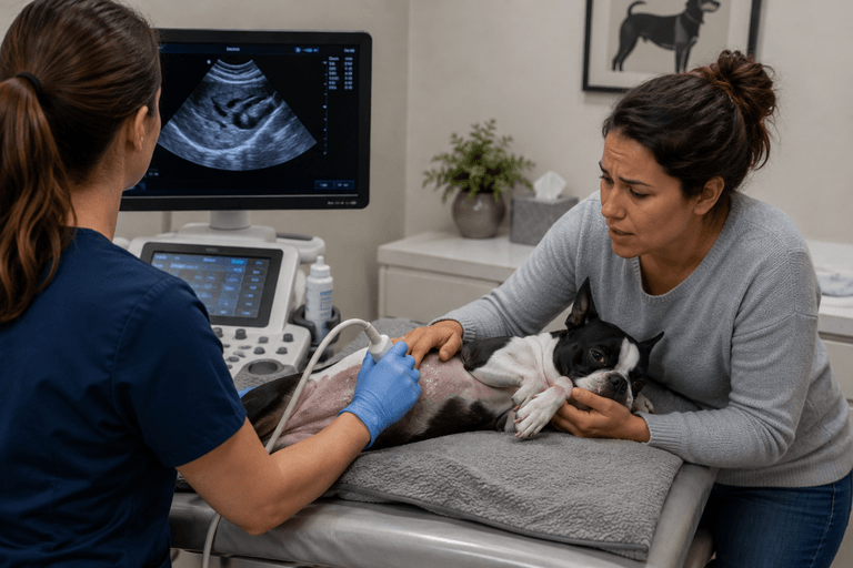
Veterinary ultrasounds and echocardiograms give veterinarians another way to investigate what is happening inside a patient’s body. Instead of relying on symptoms alone, imaging allows the veterinary team to evaluate internal structures and, in the case of echocardiography, cardiac function in real time.
At Veterinary Diagnostics Centers, we understand that the goal of diagnostic imaging isn’t simply to produce more pictures. It is to provide useful information that helps veterinarians understand complicated cases and determine what should happen next.
Some Pet Health Problems Don’t Follow a Straight Line
In an ideal diagnostic case, a pet develops a symptom, a veterinarian performs a test, and the answer becomes obvious.
Real cases are rarely that simple.
A pet may show several symptoms at once, but laboratory results can sometimes identify an abnormality that requires further investigation. A physical examination may reveal a concern that needs further investigation. Previous diagnostics may rule out some possibilities while leaving several others on the table.
This is particularly challenging because dogs and cats cannot tell us exactly what they are experiencing.
They cannot explain whether their discomfort is coming from their stomach, liver, kidneys, or another part of the body.
Instead, veterinarians have to piece together the information available to them. Advanced imaging becomes another important piece of that puzzle.

Ultrasound Can Help Connect the Dots
One of the strengths of abdominal ultrasound is that it allows veterinarians to evaluate several internal structures during the same examination.
Depending on the case, this can include the:
- Liver
- Gallbladder
- Kidneys
- Urinary bladder
- Pancreas
- Spleen
- Stomach
- Intestinal tract
- Adrenal glands
- Lymph nodes
Ultrasound provides information about internal architecture and movement in real time, offering a different type of information from radiographs.
That broader view can be especially useful when the original clinical concern doesn’t completely explain the patient’s condition.
For example, a veterinarian may be investigating an abnormal laboratory result involving one organ. During the ultrasound examination, another abnormality may be discovered elsewhere.
This does not necessarily mean the first concern was incorrect.
It may mean the patient has a more complicated medical picture than initially suspected.
Abnormal Blood Work Doesn’t Always Tell the Whole Story
Laboratory testing is an essential part of veterinary medicine.
Blood work can tell veterinarians that something isn’t functioning normally. It may identify changes involving the liver, kidneys, blood cells, electrolytes, or other aspects of a pet’s health.
But laboratory results do not always show what is causing those changes.
A pet may appear completely normal at home while routine blood work reveals an unexpected abnormality. In these situations, ultrasound can help veterinarians investigate the organs associated with the abnormal results and determine whether there are structural changes that could help explain them.
This is one of the reasons diagnostic imaging can be valuable even when a pet does not appear seriously ill.
The absence of dramatic symptoms does not necessarily mean there is nothing happening internally.
Sometimes the Most Important Finding Is Somewhere Else
One of the interesting aspects of veterinary ultrasound is that the examination can reveal something that wasn’t the original reason for referral.
The existing Veterinary Diagnostics Centers material describes situations where pets referred for one concern have unexpectedly been found to have conditions such as splenic masses, gallbladder disease, bladder stones, enlarged organs, fluid accumulation, or hidden intestinal disease.
This illustrates why a comprehensive diagnostic approach matters.
The purpose of an ultrasound isn’t to look at only the organ that appears most suspicious from the beginning.
It is an opportunity to evaluate the relevant internal structures and determine whether the imaging findings support the original clinical suspicion—or point somewhere else.
Sometimes the most valuable answer is not the one everyone expected.
When Symptoms and Imaging Tell Different Stories
Medical cases can become particularly challenging when a patient’s outward symptoms do not appear to match the initial diagnostic findings.
A pet may have significant clinical signs but relatively mild laboratory changes. Another may have substantial laboratory abnormalities with very few visible symptoms. Neither situation automatically means something is wrong with the diagnostic process. It may simply mean that additional information is needed.
Ultrasound can provide that additional perspective by allowing veterinarians to examine soft tissues and internal organs directly.
This can help determine whether the findings appear localized, whether multiple organs are involved, or whether the original suspected condition needs to be reconsidered.
The goal isn’t to force every finding into the first diagnosis.
The goal is to understand what the evidence is actually showing.
Complex Cases Often Require More Than One Diagnostic Tool
Veterinary ultrasound does not replace other diagnostics.
Instead, it complements them.
A veterinarian may use:
- Physical examination to identify clinical signs and abnormalities.
- Blood work to evaluate organ function and systemic health.
- Radiographs to examine the chest, bones, overall organ size, and other structures.
- Ultrasound to evaluate internal architecture and soft tissues in greater detail.
- Echocardiography to examine the structure and function of the heart.
Each diagnostic tool provides different information.
The attached transcript specifically emphasizes that advanced diagnostics should be used as part of the broader diagnostic process rather than automatically being the first step in every case.
That distinction is important.
Good diagnostic medicine is not about ordering every available test.
It is about selecting the tests that can provide useful information for the questions being asked.
When the Heart Becomes Part of the Bigger Picture
Some complicated cases involve more than abdominal organs.
A pet may have reduced exercise tolerance, coughing, increased respiratory effort, weakness, or another sign that raises concern about the cardiovascular system.
An echocardiogram provides a specialized way to investigate the heart.
Unlike an EKG, which evaluates the heart’s electrical activity and rhythm, an echocardiogram is a cardiac ultrasound that allows veterinarians to evaluate the physical structure and function of the heart. The transcript specifically emphasizes this distinction because the two tests are frequently confused.
An echocardiogram can provide information about:
- Heart valves
- Heart chambers
- Wall thickness
- Blood flow
- Blood flow velocity
- Direction of blood flow
- Cardiac function
This level of information can help veterinarians better understand whether a cardiac abnormality is clinically significant and what additional care may be appropriate.
A Heart Murmur Is Only One Piece of the Puzzle
A murmur can be an important reason to investigate cardiac health, but the sound itself does not explain exactly what is happening inside the heart.
The same general clinical finding can have different underlying causes.
An echocardiogram allows veterinarians to move beyond simply hearing an abnormal sound and examine the structures responsible for blood flow through the heart.
This can be especially important when a pet is being evaluated for anesthesia, showing reduced exercise tolerance, coughing, increased respiratory effort, or experiencing weakness or collapse episodes.
The goal is to understand the heart rather than simply label the finding.
Echocardiography Gives a More Detailed View of Cardiac Function
Chest X-rays can provide useful information about the overall size and shape of the heart.
An echocardiogram goes further by allowing veterinarians to evaluate internal cardiac structures and function.
The attached material identifies several areas that can be assessed through cardiac ultrasound, including valve function, wall thickness, chamber size, blood flow, pumping function, pulmonary pressures, and heart muscle disease.
This makes echocardiography particularly valuable when a veterinarian needs more information about a suspected cardiac condition.
It is not simply about determining whether the heart is “large.”
It is about understanding how the heart is built and how it is working.
Sometimes Ultrasound Helps Rule Things Out
Pet owners often think of diagnostic testing as a search for a positive diagnosis. But ruling out serious problems can be just as important.
If ultrasound does not show evidence of a suspected obstruction, mass, fluid accumulation, or another major abnormality, that information can still help guide the diagnostic process.
It may allow the veterinary team to focus on other possible causes.
The source material specifically notes that ultrasound can help veterinarians determine whether surgery is needed, whether disease appears localized or widespread, which organs are involved, whether additional testing is needed, and how urgent treatment may be.
Sometimes knowing what isn’t happening inside the body is an important part of finding out what is.
An Ultrasound Finding May Lead to the Next Question
It is also important for pet owners to understand that ultrasound does not always provide a final diagnosis.
An ultrasound may identify a mass, abnormal lymph nodes, thickened intestines, inflammation, or another structural change.
But imaging cannot always determine the exact microscopic type of disease.
For example, a mass may be visible on ultrasound, but additional testing such as fine-needle aspiration, cytology, biopsy, blood work, CT, or follow-up imaging may still be recommended.
This does not make the ultrasound less valuable.
In many cases, identifying where the problem is is a major step toward determining exactly what it is.
The attached transcript makes the same point directly: an ultrasound can reveal that the liver, for example, is the area of concern, while additional testing is needed to determine the specific disease affecting it.
The Value of Asking Better Diagnostic Questions
A complex case often becomes clearer when the veterinary team can ask a more specific question.
Instead of simply asking:
“What’s wrong with my pet?”
the diagnostic process can move toward questions such as:
- Is there structural disease in the liver?
- Is the gastrointestinal tract abnormal?
- Is there evidence of an obstruction?
- Is a mass present?
- Are multiple organs affected?
- Is the heart structurally normal?
- Is a detected murmur associated with significant cardiac disease?
- Does the imaging support the current treatment plan?
- Is additional testing necessary?
Those questions are much easier to investigate with the right diagnostic tools. This is where veterinary ultrasounds and echocardiograms become particularly useful. They help transform a broad clinical concern into specific information that veterinarians can use.
Better Information Can Lead to More Individualized Care
Every dog and cat is different.
Age, breed, medical history, symptoms, laboratory results, previous imaging, and response to treatment can all influence how a case is approached.
That means the same imaging finding may not have exactly the same significance in every patient.
Advanced diagnostic imaging helps veterinarians build treatment recommendations around the individual patient rather than relying only on general assumptions.
For some pets, imaging may support continued monitoring.
For others, it may lead to additional testing.
In another patient, it may indicate that treatment needs to be changed or that surgery should be considered.
The value is in having better information before making those decisions.
Why Real-Time Imaging Matters
One of the defining characteristics of veterinary ultrasound is that the examination happens in real time.
The sonographer can adjust the probe, change imaging angles, follow structures, and investigate findings as they appear.
The existing material explains that ultrasound is highly operator-dependent because the examination requires real-time technical decision-making.
This is particularly relevant in complicated cases.
Unexpected findings may require additional views.
An area that initially looks normal may need closer examination because of something discovered elsewhere.
The scan can evolve based on what is actually being seen.
That flexibility is one of the reasons ultrasound remains such a valuable part of advanced veterinary diagnostics.
Helping the Veterinary Team Decide What Comes Next
A diagnostic test is most useful when its findings can influence patient care.
Ultrasound and echocardiography can help veterinarians determine whether they need to:
- Continue medical management
- Change a treatment plan
- Perform additional testing
- Monitor an abnormality
- Consider surgery
- Investigate another organ system
- Evaluate cardiac function more closely
- Schedule follow-up imaging
This doesn’t mean imaging eliminates uncertainty.
Veterinary medicine will always involve some degree of uncertainty, especially in complicated cases.
But better information can make that uncertainty more manageable.
When the Answer Is More Complicated Than Expected
Sometimes an ultrasound or echocardiogram confirms what the veterinary team already suspected.
Other times, it changes the direction of the case.
That can be difficult for owners to hear, especially when they expected a straightforward answer.
But discovering that a case is more complicated is still useful information.
It allows the veterinary team to adjust the diagnostic plan rather than continuing to treat an assumption.
The goal is not simply to find an answer quickly.
It is to find the right information so that the next medical decision is based on the patient’s actual condition.
Advanced Imaging Is About Understanding the Whole Patient
Veterinary ultrasounds and echocardiograms are powerful because they allow veterinarians to investigate internal structures that cannot be fully understood from outward symptoms alone.
But their greatest value comes from placing those findings into context.
A laboratory abnormality has a story.
A symptom has a story.
A heart murmur has a story.
An ultrasound finding has a story.
An echocardiogram measurement has a story.
When those pieces are considered together, the veterinary team can develop a much clearer understanding of the patient.
At Veterinary Diagnostics Centers, advanced imaging is part of that larger diagnostic process. The purpose is to provide detailed information that helps referring veterinarians answer difficult questions, investigate complicated cases, and determine the most appropriate next steps for each individual pet.
Because sometimes the most important diagnostic breakthrough isn’t finding one obvious answer.
It is finally understanding how all the pieces fit together.