
When Test Results Don’t Tell the Whole Story: How Veterinary Ultrasounds and Echocardiograms Help Find the Missing Answers
One of the most rewarding moments in veterinary medicine is being able to give a pet owner clear answers after weeks—or even months—of uncertainty.
Many medical cases do not begin with an obvious diagnosis.
A dog may have recurring vomiting despite several rounds of medication.
A cat may lose weight while continuing to eat normally.
Routine blood work may reveal mild abnormalities that do not fully explain the symptoms a pet is experiencing.
Sometimes, everything seems almost normal on paper, yet owners know something about their pet simply is not right.
These situations can be frustrating for everyone involved.
Pet owners want to understand why their companion is not feeling well.
Veterinarians want to recommend the right treatment, but effective treatment begins with an accurate diagnosis.
This is where advanced imaging often changes the course of a case.
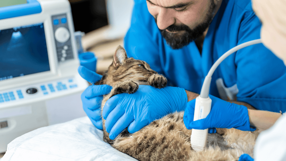
Veterinary ultrasound and echocardiography provide information that cannot be obtained through physical examinations or laboratory testing alone. They allow veterinarians to evaluate organs in real time, observe how structures function, and identify changes that may not be visible using other diagnostic tools.
Rather than replacing blood work or radiographs, ultrasound and echocardiograms complement those tests, helping veterinarians build a more complete understanding of a patient’s health.
Every Diagnostic Test Has a Different Purpose
One of the biggest misconceptions about veterinary diagnostics is that a single test should provide every answer.
In reality, each diagnostic tool contributes a different piece of the overall picture.
A physical examination helps veterinarians assess pain, mobility, body condition, heart sounds, breathing, and visible abnormalities.
Blood work evaluates organ function, infection, inflammation, and metabolic health.
Radiographs help assess bones, the lungs, and the general size and position of internal organs.
Ultrasound and echocardiography offer something entirely different.
They allow veterinarians to look inside organs, evaluate tissue characteristics, observe movement, and identify subtle structural abnormalities that cannot be appreciated through other methods.
When these diagnostic tools are interpreted together, they provide a much more complete understanding of the patient’s condition.
Why Symptoms and Laboratory Results Do Not Always Match
One of the reasons advanced imaging is so valuable is because pets do not always fit textbook descriptions.
A dog with significant liver disease may show only mild blood work changes.
A cat with intestinal disease may continue eating despite ongoing weight loss.
A pet with heart disease may have no coughing at all.
Likewise, abnormal blood work does not always indicate severe disease.
Some laboratory abnormalities are temporary.
Others represent chronic changes that require monitoring rather than immediate intervention.
The challenge is determining which findings are clinically important.
Ultrasound often provides the missing context needed to answer that question.
For example, mildly elevated liver enzymes become much more meaningful if ultrasound reveals gallbladder disease or structural changes within the liver.
Conversely, if the liver appears healthy on imaging, veterinarians may investigate entirely different causes.
This ability to connect laboratory findings with real-time anatomy is one of ultrasound’s greatest strengths.
Looking Beyond Numbers
Modern laboratory equipment provides an incredible amount of information.
However, numbers alone cannot show what an organ actually looks like.
Two dogs may have nearly identical liver enzyme values.
One may have mild inflammation.
The other may have a large mass involving the liver.
Without imaging, those two situations can appear surprisingly similar.
Ultrasound allows veterinarians to move beyond laboratory values and directly evaluate:
- Organ size
- Tissue appearance
- Internal architecture
- Blood vessels
- Fluid accumulation
- Abnormal masses
- Cysts
- Mineralization
- Lymph nodes
These findings often explain why blood work appears abnormal and help determine the most appropriate next step.
When Ultrasound Changes the Direction of a Case
Veterinarians frequently encounter cases where the original concern turns out to be only part of a larger picture.
A pet may be referred because of elevated kidney values.
During ultrasound, the kidneys appear relatively stable, but gallbladder abnormalities or intestinal disease are discovered instead.
Similarly, a dog evaluated for vomiting may unexpectedly have a splenic mass that was not suspected during the initial examination.
These unexpected findings are one reason ultrasound has become such an important diagnostic resource.
Rather than focusing on only one organ, a complete abdominal ultrasound evaluates multiple body systems during the same examination.
This comprehensive approach often identifies problems that might otherwise remain hidden.
Understanding Echocardiography Beyond the Heart Murmur
Many pet owners assume an echocardiogram is only necessary after a veterinarian hears a heart murmur.
While murmurs are certainly one of the most common reasons for referral, they are not the only reason cardiac imaging is recommended.
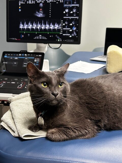
An echocardiogram evaluates how the heart functions—not simply how it sounds.
This distinction is important because some pets with significant heart disease have only subtle murmurs, while others with louder murmurs may have relatively mild structural changes.
Cardiac ultrasound allows veterinarians to evaluate:
- Heart chamber size
- Valve function
- Blood flow through the heart
- Heart muscle thickness
- Pumping efficiency
- Congenital abnormalities
- Pulmonary pressures
These measurements provide objective information that cannot be obtained with a stethoscope alone.
Why Earlier Answers Often Mean Better Planning
One of the greatest benefits of advanced imaging is not simply identifying disease.
It is identifying disease early enough to make informed medical decisions.
Whether the diagnosis ultimately involves medication, surgery, dietary management, or ongoing monitoring, having accurate information allows veterinarians and pet owners to make decisions with greater confidence.
Early imaging may also help avoid unnecessary treatments.
Rather than trying multiple medications without understanding the underlying cause, veterinarians can tailor recommendations based on what imaging reveals.
This more targeted approach often improves efficiency while reducing uncertainty for everyone involved.
Advanced Imaging Supports Better Communication
Another overlooked advantage of ultrasound and echocardiography is how they improve communication between veterinary teams and pet owners.
Being able to visualize an organ or explain heart function using real-time imaging often helps owners better understand their pet’s condition.
Instead of discussing possibilities in general terms, veterinarians can explain what they are seeing and how those findings relate to symptoms, laboratory results, and treatment recommendations.
This collaborative approach allows owners to make informed decisions while feeling more confident about the diagnostic process.
When Chronic Conditions Need More Than Routine Monitoring
Not every medical condition can be cured.
Many diseases affecting dogs and cats are managed over months or years, making ongoing monitoring an important part of long-term care.
Veterinary ultrasound plays a valuable role because it allows veterinarians to compare changes over time rather than relying solely on symptoms.
A pet may appear stable at home, yet imaging can reveal gradual progression that deserves attention before noticeable clinical signs develop.
Repeat ultrasound examinations are commonly recommended for patients with:
- Chronic liver disease
- Gallbladder abnormalities
- Kidney disease
- Splenic nodules
- Pancreatic disease
- Bladder masses
- Intestinal disorders
- Certain benign growths that require observation
Monitoring these conditions allows veterinarians to determine whether treatment is working, whether medications should be adjusted, or whether additional diagnostics have become necessary.
Instead of reacting after significant deterioration occurs, imaging supports a proactive approach to patient care.
Ultrasound Can Help Clarify Unexpected Findings
Veterinary medicine rarely follows a predictable path.
Some of the most valuable ultrasound examinations are those that uncover findings no one expected.
For example, a dog may be referred because of mild elevations in liver enzymes.
During the examination, the liver appears relatively normal, but the gallbladder shows significant abnormalities that explain the laboratory changes.
In another case, a cat being evaluated for chronic weight loss may have normal intestinal architecture but enlarged abdominal lymph nodes that warrant additional investigation.
Sometimes the opposite occurs.
An abnormal blood test may appear concerning, yet ultrasound demonstrates healthy organs with no structural abnormalities.
That information can be equally important because it helps veterinarians avoid unnecessary procedures while continuing to investigate other possible causes.
Rather than confirming assumptions, ultrasound helps veterinarians follow the evidence wherever it leads.
The Difference Between Seeing an Organ and Understanding It
Many people think ultrasound simply produces pictures.
In reality, diagnostic ultrasound is a dynamic examination.
Every movement of the probe provides additional information.
The sonographer evaluates how organs interact with one another, how tissues respond to pressure, and whether subtle abnormalities become visible from different angles.
This process requires continuous interpretation.
During a complete abdominal ultrasound, veterinarians assess:
- Overall organ size
- Internal tissue patterns
- Organ borders
- Blood vessel appearance
- Fluid accumulation
- Presence of cysts or masses
- Changes within lymph nodes
- Relationships between surrounding organs
Each observation contributes another piece to the diagnostic puzzle.
This is one reason ultrasound remains one of the most valuable imaging tools available in veterinary medicine.
Why Echocardiograms Are About More Than Diagnosing Heart Disease
Heart disease is certainly one of the primary reasons veterinarians recommend echocardiography.
However, cardiac ultrasound also helps answer many questions that extend beyond the heart itself.
Understanding cardiovascular function may influence decisions involving:
- Dental procedures requiring anesthesia
- Orthopedic surgery
- Emergency procedures
- Long-term medication planning
- Exercise recommendations
- Monitoring chronic respiratory conditions
A pet with previously undiagnosed heart disease may require a very different anesthetic protocol than one with normal cardiac function.
Likewise, understanding how efficiently the heart pumps blood helps veterinarians determine whether fatigue, weakness, or exercise intolerance is related to cardiac disease or another medical condition.
The information gained through echocardiography often supports safer treatment planning across many areas of veterinary medicine.
When Experience Makes the Difference
Modern ultrasound equipment continues to improve every year.
Higher image resolution, better Doppler technology, and enhanced processing capabilities have made veterinary imaging more powerful than ever before.
Yet technology is only one part of the diagnostic process.
Ultrasound remains one of the most operator-dependent imaging modalities in veterinary medicine.
The quality of the examination depends heavily on the experience of the person performing and interpreting the scan.
Experienced sonographers recognize subtle changes that may be overlooked during less comprehensive evaluations.
They know when an organ appears normal, when a finding deserves closer investigation, and when additional diagnostic procedures may provide valuable information.
That expertise becomes especially important when evaluating small lesions, complex abdominal disease, or early cardiac abnormalities.
High-quality equipment and experienced interpretation work together to provide the most accurate diagnostic information possible.
Advanced Imaging Supports Collaborative Veterinary Medicine
Today’s veterinary care often involves collaboration between multiple professionals.
Primary care veterinarians, emergency clinicians, internal medicine specialists, surgeons, cardiologists, and diagnostic imaging providers frequently work together to manage complex medical cases.
Ultrasound findings become an important part of this collaborative process.
Detailed imaging reports help referring veterinarians better understand the patient’s condition while supporting informed discussions with pet owners.
This team-based approach allows every provider involved in the patient’s care to make recommendations based on the same comprehensive diagnostic information.
Whether a pet ultimately requires surgery, medical management, additional laboratory testing, or long-term monitoring, advanced imaging often provides the foundation for those decisions.
Better Answers Lead to Better Care
Veterinary medicine is built on careful observation, thoughtful diagnostics, and individualized treatment planning.
Every pet presents a unique combination of history, symptoms, physical examination findings, and diagnostic results.
Veterinary ultrasound and echocardiography help connect those pieces together.
They provide insight into organ health, cardiac function, and disease progression that cannot be obtained through physical examination alone.
For pets experiencing unexplained illness, persistent symptoms, abnormal laboratory findings, or suspected heart disease, advanced imaging frequently provides the clarity needed to move forward with confidence.
The ultimate goal is not simply to perform another diagnostic test.
It is to understand each patient more completely so veterinarians can recommend care that is accurate, timely, and tailored to that individual pet.