
Why Timing Matters: When Veterinary Ultrasounds and Echocardiograms Can Make the Biggest Difference
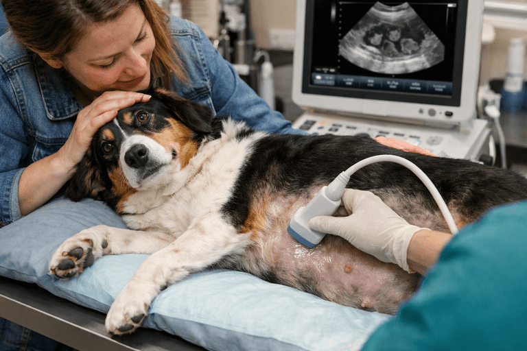
One of the most challenging aspects of veterinary medicine is determining when advanced diagnostics should become part of a pet’s care plan.
Every veterinarian has experienced cases where a pet’s symptoms seem mild at first, only to discover later that a more significant condition has been developing beneath the surface.
A dog with occasional vomiting may actually have gallbladder disease.
A cat with subtle weight loss may have intestinal changes that warrant further investigation.
A heart murmur detected during a routine wellness exam may indicate early cardiac disease that is not yet causing noticeable symptoms at home.
The difficulty is that pets rarely tell us when something is wrong.
Unlike people, they cannot explain where they hurt, describe changes in their energy level, or tell us when symptoms first began. Many animals instinctively hide signs of illness until a condition has progressed.
Because of this, timing plays an important role in veterinary diagnostics.
Knowing when to pursue advanced imaging can help veterinarians identify disease earlier, guide treatment decisions more effectively, and improve outcomes for pets.
Veterinary ultrasounds and echocardiograms are among the most valuable diagnostic tools available because they allow veterinarians to look beyond symptoms and evaluate what is happening inside the body in real time.
Why Waiting for Symptoms to Worsen Can Delay Diagnosis
Many pet owners assume that severe symptoms are necessary before advanced imaging is recommended.
In reality, some of the most important findings occur when symptoms are still subtle.
Conditions involving the liver, gallbladder, pancreas, kidneys, intestines, and heart often develop
gradually. Early signs may be easy to overlook or may seem unrelated.
Examples include:
- Mild decreases in appetite
- Intermittent vomiting
- Changes in water consumption
- Weight loss despite normal eating habits
- Reduced activity levels
- Exercise intolerance
- Occasional coughing
- Changes in urinary habits
These symptoms can have many causes, making it difficult to determine what is happening through observation alone.
In many cases, blood work and physical examination identify that something is abnormal but cannot pinpoint the source of the problem.
Advanced imaging helps bridge that gap.
Understanding the Role of Veterinary Ultrasound
Veterinary ultrasound uses sound waves to create real-time images of internal organs and soft tissues.

Unlike X-rays, which primarily show organ size and shape, ultrasound allows veterinarians to evaluate internal architecture, tissue characteristics, blood flow, and organ movement.
Ultrasound is commonly used to assess:
- Liver
- Gallbladder
- Pancreas
- Kidneys
- Bladder
- Spleen
- Intestinal tract
- Lymph nodes
- Adrenal glands
One of the greatest benefits of ultrasound is that it provides detailed information without exposing pets to radiation.
For many patients, ultrasound becomes the next step when physical examinations, blood tests, and radiographs do not provide enough information to explain a pet’s symptoms.
When Blood Work Raises Questions
Routine laboratory testing often provides the first indication that something may be wrong.
Elevated liver enzymes, kidney value changes, abnormal pancreatic markers, or unexplained anemia can all suggest underlying disease.
However, blood work rarely tells the entire story.
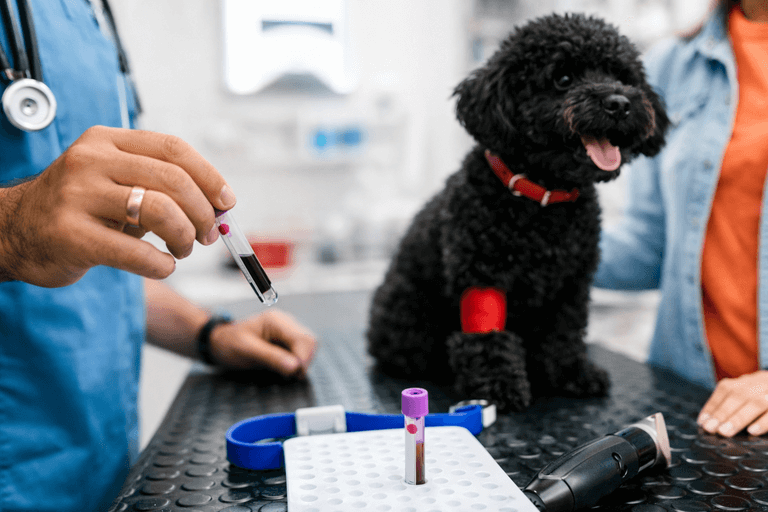
Abnormal values indicate that something is happening, but they do not always reveal which organ is affected or what condition is causing the changes. For example, elevated liver values do not always mean the liver itself is the problem.
Gallbladder disease, pancreatic inflammation, intestinal disorders, and systemic illness can all influence liver enzymes.
Ultrasound helps veterinarians evaluate the organs involved and determine which findings are clinically significant.
This information can substantially influence treatment recommendations and next steps.
Gastrointestinal Symptoms Often Require More Than Basic Diagnostics
Vomiting, diarrhea, appetite changes, and weight loss are among the most common reasons veterinarians recommend abdominal ultrasound.
These symptoms can result from a wide range of conditions, including:
- Inflammatory bowel disease
- Pancreatitis
- Gallbladder disease
- Intestinal foreign bodies
- Gastrointestinal tumors
- Liver disease
- Kidney disease
Many of these conditions produce similar symptoms, making diagnosis challenging without advanced imaging.
Ultrasound allows veterinarians to evaluate intestinal wall thickness, identify abnormal lymph nodes, assess surrounding organs, and detect changes that may not appear on X-rays.
This is especially important for cats, which often show subtle signs of illness until disease has progressed significantly.
The Importance of Ultrasound for Urinary Conditions
Urinary problems can become emergencies quickly.
Straining to urinate, blood in the urine, increased urinary frequency, or accidents in previously house-trained pets should never be ignored.
Ultrasound can help identify:
- Bladder stones
- Kidney abnormalities
- Urinary obstruction
- Bladder masses
- Chronic inflammation
In cases involving urinary blockage, rapid diagnosis is essential because delayed treatment can become life-threatening.
Advanced imaging helps veterinarians determine the cause of urinary symptoms and whether immediate intervention is necessary.
Why Heart Murmurs Should Not Be Ignored
Heart murmurs are among the most common reasons veterinarians recommend an echocardiogram.
A murmur is the sound of turbulent blood flow within the heart.
However, a murmur alone does not explain why that turbulence is occurring. Some murmurs are relatively benign. Others indicate significant structural heart disease.
An echocardiogram allows veterinarians to evaluate the heart directly and determine the cause and severity of the problem.
What an Echocardiogram Reveals
An echocardiogram is a specialized ultrasound that evaluates the heart in motion.
Unlike chest X-rays, which show only the heart’s overall shape and size, echocardiography provides detailed information about cardiac function.
Veterinarians can evaluate:
- Heart valve function
- Chamber size
- Blood flow direction
- Heart muscle thickness
- Pumping efficiency
- Pulmonary pressures
This information is essential for diagnosing and managing heart disease.
In dogs, echocardiograms commonly identify degenerative valve disease.
In cats, they frequently detect cardiomyopathy, a condition involving abnormal thickening of the heart muscle.
Early identification helps veterinarians monitor disease progression and determine when
treatment is appropriate.
Situations Where Earlier Imaging Can Change Outcomes
There are many cases in which earlier ultrasound or echocardiography can significantly alter a pet’s care plan.
Examples include:
- Detecting a splenic mass before rupture occurs
- Identifying gallbladder disease before complications develop
- Diagnosing heart disease before congestive heart failure develops
- Finding urinary obstruction before kidney damage occurs
- Monitoring tumors before they become symptomatic
- Detecting intestinal abnormalities before severe weight loss occurs
Early diagnosis does not guarantee a cure, but it often creates more treatment options and allows veterinarians to intervene before a condition becomes critical.
Why Experience Is Essential in Veterinary Imaging
Ultrasound is one of the most operator-dependent imaging modalities in veterinary medicine.
Unlike CT scans or MRI studies, ultrasound requires real-time decision-making throughout the examination. A skilled sonographer does more than capture images.
They interpret findings as they scan, adjust techniques based on patient anatomy, and investigate subtle abnormalities that may otherwise be overlooked.
Experience is especially important when evaluating:
- Intestinal disease
- Gallbladder abnormalities
- Early tumors
- Small structures such as adrenal glands
- Cardiac function
High-quality equipment matters, but expertise remains equally important. The combination of advanced technology and experienced interpretation helps ensure the most accurate results possible.
How Ultrasound and Echocardiography Support Long-Term Care
Advanced imaging is not only useful for diagnosing new conditions. It also plays an important role in monitoring chronic disease.
Repeat ultrasounds and echocardiograms may be recommended to:
- Monitor heart disease progression
- Evaluate treatment effectiveness
- Track changes in tumors
- Reassess gallbladder disease
- Monitor chronic kidney disease
- Follow intestinal abnormalities over time
Comparing imaging studies over months or years helps veterinarians recognize subtle changes that may influence treatment decisions.
This proactive approach allows care plans to evolve alongside a pet’s needs.
The Value of Finding Answers Earlier
Pet owners often hope that symptoms will improve on their own. Sometimes they do.
Other times, delayed evaluation can allow disease to progress unnoticed.
Advanced imaging is not necessary for every patient, but knowing when to pursue ultrasound or echocardiography can make a meaningful difference.
These tools help veterinarians identify hidden disease, clarify confusing symptoms, and develop treatment plans based on a more complete understanding of what is happening inside the patient.
For pets with unexplained symptoms, abnormal blood work, urinary concerns, heart murmurs, or chronic digestive issues, advanced imaging often provides the information needed to move forward with greater confidence.