
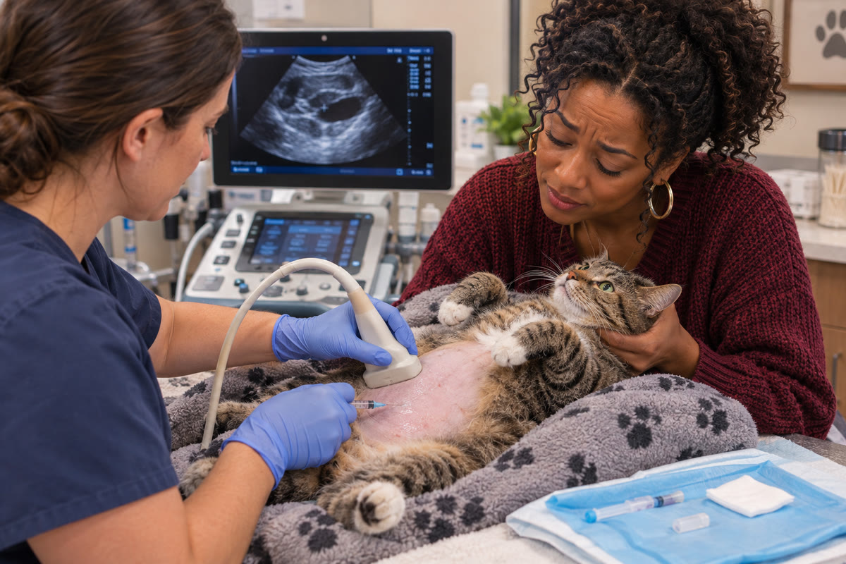
A pet's abdomen can become noticeably larger for several reasons. Weight gain, digestive changes, organ enlargement, and fluid accumulation can all affect the shape of the belly. Although these changes may look similar from the outside, they can have very different causes and require different approaches to diagnosis.
One possible explanation is abdominal effusion, which occurs when fluid accumulates within the abdominal cavity. The fluid may develop because of problems affecting the liver, heart, blood vessels, abdominal organs, or the lining of the abdominal cavity. In some cases, it may also be associated with inflammation, injury, or internal bleeding.
Because several conditions can produce similar outward signs, determining the cause requires more than looking at the size of a pet's abdomen. A veterinarian must evaluate the animal's overall condition and determine which diagnostic tests can provide the most useful information.
Veterinary ultrasounds and echos can play important roles in this process. An abdominal ultrasound allows a veterinary professional to examine internal organs and identify fluid within the abdomen. An echocardiogram, also called an echo, evaluates the heart's structure and function when cardiovascular disease is a possible contributor.
These examinations answer different questions, but their findings can help build a clearer picture of what is happening inside a pet's body. Understanding how each test contributes to the diagnostic process can help pet owners better understand why additional imaging may be recommended.
What Does Fluid in a Pet's Abdomen Mean?
The medical term for fluid accumulation within the abdominal cavity is ascites. More broadly, abdominal effusion refers to fluid that collects in the space surrounding the abdominal organs.
A small amount of fluid may be difficult to recognize during an ordinary physical examination. As more fluid accumulates, the abdomen may become enlarged or appear rounded. Some pets develop discomfort, reduced activity, or changes in appetite. Others may show signs associated with the underlying disease rather than obvious abdominal swelling.
The presence of fluid is a finding, not a complete diagnosis.
For example, two dogs may both have enlarged abdomens, but one may have fluid related to cardiovascular disease while the other may have fluid associated with abdominal inflammation or internal bleeding. The appearance of the abdomen alone cannot reliably distinguish between these possibilities.
The characteristics of the fluid also matter. Fluid can differ in its protein content, cellular composition, and other measurable properties. These differences may help veterinarians narrow the list of possible causes.
However, the fluid must be interpreted alongside the pet's medical history, physical examination, blood test results, and imaging findings. No single observation necessarily explains the entire condition.
This is where diagnostic imaging becomes valuable. Rather than simply confirming that the abdomen looks enlarged, ultrasound can help determine whether fluid is present, show how it relates to surrounding structures, and identify abnormalities that may guide further investigation.

Why an Abdominal Ultrasound Can Be Important
An abdominal ultrasound uses high-frequency sound waves to create real-time images of internal structures. Unlike an examination that relies only on external signs, ultrasound allows a trained veterinary professional to evaluate organs and tissues within the abdomen.
When fluid accumulation is suspected, the examination can help answer several important questions:
- Is fluid present within the abdominal cavity?
- How is the fluid distributed around the organs?
- Are there visible abnormalities involving the liver, spleen, kidneys, bladder, or gastrointestinal tract?
- Are any organs enlarged or showing changes in their internal appearance?
- Is there a mass or another abnormality that requires further investigation?
- Could the findings help determine whether fluid sampling or additional testing is needed?
These questions matter because abdominal fluid may occur alongside changes in the organs or tissues responsible for maintaining normal fluid balance.
For example, an ultrasound may identify changes involving the liver or show an abdominal mass that warrants further investigation. In another patient, the organs may have no obvious focal abnormality, making blood tests, fluid analysis, cardiovascular evaluation, and other diagnostic information especially important.
Ultrasound also allows the examiner to evaluate structures from multiple angles during the same appointment. If an area appears abnormal, the sonographer can adjust the imaging view to investigate it more closely.
This real-time assessment is particularly useful when the initial findings are unexpected or when the pet's symptoms point toward a different part of the abdomen than the area where an abnormality is discovered.
Importantly, identifying fluid does not automatically establish why it developed. The ultrasound provides anatomical information that must be interpreted within the wider diagnostic picture.
How the Location and Appearance of Fluid Guide the Investigation
Fluid may collect in different areas of the abdominal cavity, and its distribution can influence what the veterinary team investigates next.
Ultrasound allows the examiner to observe fluid in relation to the abdominal organs. It may also reveal changes such as tissue thickening, irregular organ contours, enlarged structures, or material within the fluid.
These observations help direct attention toward areas that deserve closer examination.
For instance, fluid accompanied by abnormalities involving the liver may prompt further evaluation of liver function and circulation. Fluid associated with visible changes in the intestinal tract may raise concern for an inflammatory or other gastrointestinal process. If an abdominal mass is present, the veterinarian may need to investigate its origin and determine whether it is affecting surrounding structures.
The appearance of the fluid can provide additional clues, but ultrasound cannot always determine its exact composition. Blood, inflammatory fluid, and other types of effusion cannot be reliably distinguished in every case by appearance alone.
When appropriate, a veterinarian may recommend collecting a fluid sample for laboratory analysis. Examining the sample can provide information about its cells, protein content, and other characteristics. Depending on the findings, additional testing may include cytology, culture, or specific biochemical measurements.
This combination of imaging and laboratory analysis is important because a visible abnormality does not always explain the full clinical picture.
A mass, for example, may be associated with fluid accumulation without ultrasound being able to determine whether the mass is benign or malignant. Further testing may be needed to establish its nature.
Similarly, the presence of fluid does not necessarily mean that an organ has ruptured or that cancer is present. The goal is to identify the most likely explanation using the available evidence rather than drawing conclusions from a single finding.
When the Heart May Be Involved
Although an enlarged abdomen often leads to an investigation of the abdominal organs, the heart can also be relevant.
Certain forms of cardiovascular disease can increase pressure within the veins returning blood to the heart. When this pressure becomes abnormal, fluid may accumulate in tissues or body cavities. In some dogs, right-sided heart failure can contribute to ascites.
However, not every pet with abdominal fluid has heart disease. Liver disorders, low blood protein levels, inflammation, bleeding, and other conditions can also cause fluid accumulation.
A physical examination may identify findings that raise suspicion of a cardiovascular problem. Depending on the patient, these may include abnormal heart sounds, changes in pulse quality, reduced exercise tolerance, weakness, or other signs of circulatory dysfunction.
Even so, a physical examination alone may not establish whether the heart is responsible for the fluid buildup.
This is one reason a veterinarian may recommend an echocardiogram when cardiovascular disease is suspected.
An echocardiogram uses ultrasound technology specifically to evaluate the heart. It allows the examiner to assess the heart's chambers, valves, muscle walls, movement, and blood flow. Doppler techniques can provide additional information about the direction and velocity of blood moving through the heart and major vessels.
These measurements help the veterinary team investigate whether structural or functional heart abnormalities could explain the pet's clinical signs.
The findings must still be considered alongside the abdominal ultrasound, physical examination, laboratory results, and overall condition of the animal. An abnormal echocardiogram does not automatically prove that heart disease is the cause of abdominal fluid. The veterinarian must determine whether the cardiac findings are severe and relevant enough to explain the problem.
Likewise, a normal or relatively reassuring echocardiogram may lead the team to investigate noncardiac causes more closely.
What an Echocardiogram Can Reveal That an Abdominal Ultrasound Cannot
Both abdominal ultrasound and echocardiography use sound waves, but they are designed to answer different diagnostic questions.
An abdominal ultrasound focuses on organs and tissues within the abdomen. An echocardiogram focuses on the heart and its function.
An abdominal scan may reveal fluid surrounding the liver or other organs, but it does not provide the same detailed assessment of cardiac chamber dimensions, valve movement, and blood flow that a dedicated echocardiogram can provide.
Conversely, an echocardiogram can reveal heart abnormalities that may help explain a pet's circulatory problems, but it does not replace a comprehensive abdominal examination when the cause of swelling remains uncertain.
Consider a dog that develops a progressively enlarged abdomen along with reduced stamina. An abdominal ultrasound may confirm fluid accumulation and evaluate the liver, spleen, and other abdominal structures. If the history and examination raise concern about cardiovascular disease, an echocardiogram may then be recommended to evaluate the heart.
The two examinations contribute different information. Together, their findings may help the veterinarian distinguish a primarily abdominal problem from one involving the cardiovascular system.
Another pet may have abdominal fluid and a visible liver abnormality. In that situation, the diagnostic priority may be investigating the liver and determining whether the fluid is related to liver disease or another process. An echocardiogram may be useful if other findings suggest a cardiac contribution, but it is not automatically necessary for every patient with ascites.
The decision to perform one or both examinations depends on the clinical question. The most useful diagnostic plan is the one that addresses the pet's specific findings rather than applying the same collection of tests to every patient.
Why Blood Tests and Fluid Analysis Still Matter
Ultrasound provides valuable anatomical information, but it cannot measure every aspect of organ function.
Blood tests can help evaluate factors such as liver and kidney function, blood cell counts, protein concentrations, and other indicators of illness. Depending on the findings, these tests may reveal abnormalities that are not fully explained by the ultrasound images.
For example, low blood protein levels can contribute to fluid accumulation because proteins help maintain the balance of fluid between blood vessels and surrounding tissues. A blood test may help identify this problem, while ultrasound can show whether fluid has accumulated and whether visible organ abnormalities are also present.
Other blood test findings may point toward inflammation, blood loss, or an underlying organ disorder. Their significance depends on the patient's overall condition and the specific pattern of abnormalities.
Fluid analysis can add another layer of information. If a veterinarian collects abdominal fluid, laboratory evaluation may help determine whether the sample contains substantial numbers of inflammatory cells, blood, microorganisms, or other findings that warrant further investigation.
In selected cases, comparing fluid measurements with blood results can help narrow the possible causes.
Not every pet needs fluid sampling. The decision depends on the amount of fluid present, the animal's stability, the suspected cause, and whether obtaining a sample is likely to change the diagnostic plan.
When sampling is appropriate, ultrasound may help the veterinary professional identify a suitable location for collection. This can be particularly useful when fluid is distributed unevenly or when nearby structures need to be avoided.
The purpose of combining these tests is not to collect as much information as possible without direction. It is to obtain the information needed to distinguish between plausible causes and determine the next appropriate step.
What Happens When Ultrasound Finds an Abnormality?
An abdominal ultrasound may produce several different types of findings.
In some cases, the scan identifies a recognizable abnormality that strongly supports a particular diagnosis. In others, it reveals a change that narrows the possibilities but does not establish the cause.
For example, an ultrasound may show an enlarged organ, an irregular area of tissue, enlarged lymph nodes, or a mass. These findings can help identify where a problem is located, but they do not always reveal the exact disease responsible.
A mass may require additional evaluation to determine its tissue type. Depending on its location and appearance, the veterinarian may recommend fine-needle aspiration, biopsy, cytology, or another diagnostic procedure.
Similarly, changes involving the intestinal wall may be associated with several different conditions. Ultrasound can help identify the location and pattern of the abnormality, but additional testing may be necessary to distinguish among inflammatory disease, infiltrative conditions, and other causes.
If the scan does not identify an obvious source of fluid, the investigation may continue through blood tests, fluid analysis, echocardiography, or other appropriate examinations.
This does not mean the ultrasound has failed. A useful diagnostic examination can eliminate some possibilities, identify complications, or establish which questions need to be answered next.
The findings may also affect how urgently a pet needs treatment. Evidence suggesting internal bleeding, severe inflammation, organ injury, or another serious problem can prompt immediate clinical assessment.
The veterinarian must interpret the images in context and communicate which findings are significant, which remain uncertain, and whether further testing is recommended.

Recognizing When Abdominal Swelling Needs Urgent Attention
A gradually enlarging abdomen should not be ignored, particularly when the cause is unknown. However, sudden abdominal enlargement or swelling accompanied by signs of serious illness warrants urgent veterinary evaluation.
Seek emergency veterinary care if your pet develops abdominal swelling alongside signs such as:
- Difficulty breathing or unusually rapid, labored breathing
- Collapse, fainting, or profound weakness
- Pale or unusually discolored gums
- Sudden severe abdominal pain
- Marked lethargy or reduced responsiveness
- Repeated unproductive retching, especially with a rapidly enlarging abdomen
- Sudden deterioration after an injury
These signs can occur with several serious conditions, and they do not all indicate abdominal fluid. For example, acute abdominal distention may also occur with gastric dilatation-volvulus, a life-threatening condition that requires immediate treatment.
Do not wait for a scheduled imaging appointment if your pet is showing signs of an emergency. Stabilization and prompt veterinary assessment take priority, and the veterinary team will determine which diagnostic tests are appropriate.
In an emergency, ultrasound may help identify fluid or other abnormalities quickly, depending on the patient's condition and the available equipment and expertise. More comprehensive imaging may follow once the pet is stable enough for further evaluation.
The urgency of the situation is determined by the pet's clinical condition, not simply by whether an ultrasound can be performed.
Why the Experience of the Ultrasound Examiner Matters
Ultrasound is a real-time examination. The quality of the diagnostic information depends not only on the equipment but also on how the examiner obtains and interprets the images.
The person performing the examination must identify the relevant structures, adjust the probe angle, assess different views, and recognize abnormalities that may be subtle or difficult to visualize.
This matters when evaluating abdominal fluid because the underlying cause may not be obvious. A careful examination may reveal an abnormality involving an organ or tissue that would otherwise receive less attention.
The presence of fluid can sometimes improve the visibility of certain structures, but it does not make every part of the abdomen easy to assess. Intestinal gas, patient movement, body conformation, and the location of an abnormality can affect image quality.
A thorough examination also requires the findings to be interpreted alongside the reason for the referral. When the clinical question concerns possible liver disease, for example, the examiner must evaluate relevant structures while remaining attentive to unexpected abnormalities elsewhere.
Echocardiography presents its own technical demands. Evaluating the heart requires appropriate imaging views and measurements that account for the animal's size, heart rate, and other relevant factors. Blood-flow measurements must also be interpreted in the context of the heart's anatomy and function.
For both types of imaging, the report should explain the significant findings and any limitations that affect interpretation.
The aim is not simply to produce images. It is to obtain useful diagnostic information that helps the veterinary team decide what the findings mean for the individual patient.
What Pet Owners Can Expect During the Diagnostic Process
When a veterinarian recommends an abdominal ultrasound, an echocardiogram, or both, the appointment generally begins with a review of the pet's medical history and current clinical findings.
The veterinary team may ask when the abdominal swelling began, whether it has changed, and whether the pet has experienced changes in appetite, activity, breathing, urination, or bowel movements. Previous blood tests, radiographs, medications, and other diagnostic results may also be reviewed.
Preparation depends on the examination and the patient's needs. For some abdominal ultrasounds, fasting may be recommended to improve visualization of certain structures. Owners should follow the instructions provided by the veterinary team rather than withholding food or water without guidance.
During an abdominal ultrasound, the pet is positioned so the examiner can access the areas being evaluated. A small area of fur may need to be clipped, and ultrasound gel is applied to help transmit sound waves between the probe and the skin.
Many pets tolerate ultrasound without sedation. However, the approach may need to be adjusted if an animal is very anxious, painful, or unable to remain still. Any decision about sedation depends on the patient's condition and the procedure being performed.
An echocardiogram involves imaging the heart through the chest. The pet is positioned to allow the examiner to obtain the necessary views and measurements. The procedure is generally noninvasive, although the time required can vary according to the complexity of the examination and the animal's ability to cooperate.
After the examination, the findings are reviewed and communicated to the referring veterinarian or clinical team. Depending on the results, the next step may involve treatment, additional laboratory testing, fluid analysis, further imaging, or monitoring.
Not every pet with abdominal fluid requires every possible test. The diagnostic process should be tailored to the individual animal and adjusted as new information becomes available.
How Ultrasounds and Echos Help Guide the Next Decision
When fluid accumulates in a pet's abdomen, identifying the fluid is only the beginning of the investigation. The veterinary team must determine why it developed, whether the pet is stable, and which findings require immediate attention.
An abdominal ultrasound helps examine the organs and tissues within the abdomen, identify fluid, and look for abnormalities that may explain the swelling. An echocardiogram provides a more detailed evaluation of the heart when cardiovascular disease may be involved.
Blood tests and fluid analysis can help answer questions that imaging alone cannot resolve. Additional procedures may be necessary when the findings suggest a mass, inflammation, internal bleeding, or another condition that requires more specific investigation.
For pet owners, the important distinction is that an imaging finding is not always the same as a confirmed diagnosis. A scan may identify a problem, narrow the possibilities, or show that further testing is needed. Each of these outcomes can help move the diagnostic process forward.
When veterinarians select the appropriate tests and interpret the results together, they can make more informed decisions about the care a pet needs. The goal is to understand the cause of the fluid accumulation as accurately as possible and use that information to guide the next step in treatment.