Abnormal liver enzymes, recurring vomiting, jaundice, and unexplained abdominal discomfort can raise important questions about a dog's or cat's health. Although these signs may appear unrelated, they can sometimes involve the liver, gallbladder, pancreas, or structures surrounding these organs.
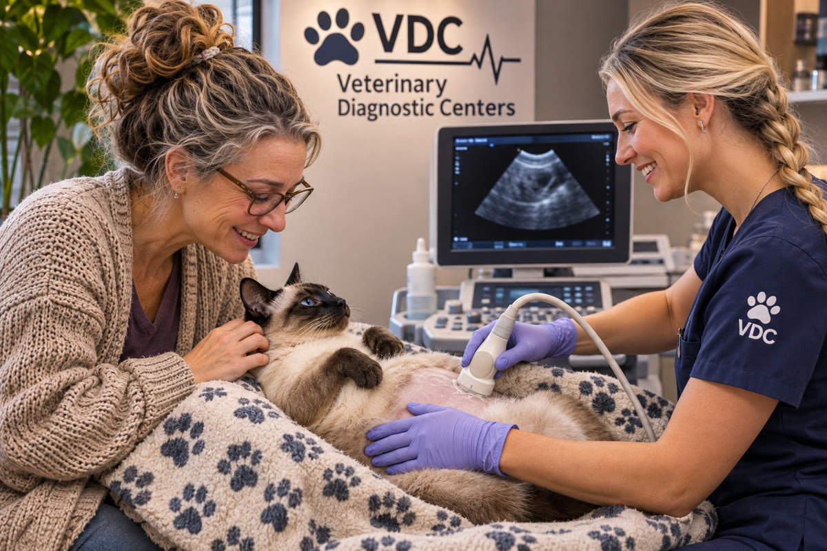
Veterinary ultrasounds help investigate these concerns by showing internal anatomy in real time. A veterinarian can examine organ size, tissue patterns, bile ducts, visible masses, surrounding fat, and fluid within the abdomen. In selected cases, ultrasound can also guide the collection of samples for laboratory analysis.
The challenge is that detecting an abnormality is not the same as identifying its exact cause. Several diseases can produce similar ultrasound appearances, and some conditions cause substantial clinical changes before obvious structural abnormalities develop.
Understanding what an ultrasound can establish—and what still requires further testing—helps explain how imaging contributes to a more complete diagnosis.
What Makes Abdominal Ultrasound Useful for These Organs?
The liver, gallbladder, and pancreas have different functions, but their anatomical locations and clinical relationships make them important parts of an abdominal diagnostic examination.
The liver processes nutrients, produces proteins, regulates aspects of metabolism, and produces bile. The gallbladder stores and concentrates bile before releasing it into the digestive tract. The pancreas produces digestive enzymes and hormones, including insulin, which helps regulate blood glucose.
An abdominal ultrasound can examine these organs alongside other structures, including the intestines, abdominal lymph nodes, blood vessels, and surrounding tissues.
This broader assessment matters because an abnormality may not originate in the organ that first appears to be involved. For example, an enlarged bile duct can prompt investigation of the gallbladder, pancreas, and other structures along the biliary system. Vomiting may justify examining the pancreas, but the intestines and other abdominal organs may also need evaluation.
Veterinarians use the clinical history and initial test results to determine which findings are most relevant. The examination should address the reason for the referral rather than simply produce a list of visible abnormalities.
The Merck Veterinary Manual's overview of ultrasonography explains that ultrasound can detect changes in organ size, shape, and tissue appearance, but many abnormalities are not specific to a single disease.

Reading Liver Ultrasound Findings in Context
The liver can develop diffuse changes that affect much of the organ or focal changes confined to particular areas. Distinguishing these patterns helps veterinarians determine which possible diagnoses deserve further investigation.
Diffuse changes versus focal lesions
A diffuse abnormality affects a broad area of the liver. On ultrasound, the tissue may appear more echogenic, less echogenic, or less uniform than expected.
A focal lesion, by contrast, is a localized area that differs from the surrounding tissue. It may be described as a nodule, mass, cyst-like structure, or another focal abnormality.
This distinction helps organize the diagnostic process, but neither pattern establishes a specific disease. Diffuse changes can occur with several metabolic, inflammatory, infiltrative, or other conditions. Focal lesions may represent benign changes, cysts, inflammation, or neoplasia.
The veterinarian considers the distribution of the changes, their appearance, the rest of the abdominal examination, and the pet's clinical findings. A lesion's size and location may influence whether sampling or additional imaging is warranted.
A normal-appearing liver also does not exclude every liver disorder. Some diseases alter cellular function or microscopic tissue architecture without producing changes that are readily visible on ultrasound.
Why liver enzyme elevations need careful interpretation
Bloodwork and ultrasound answer different diagnostic questions.
Liver-associated enzymes, such as alanine aminotransferase (ALT) and alkaline phosphatase (ALP), can increase for different reasons. ALT is commonly associated with leakage from injured liver cells, while ALP can increase with changes involving bile flow and other processes. The significance of an abnormal value depends on the species, magnitude and pattern of change, medication history, and other findings.
These enzymes do not directly measure every aspect of liver function. Veterinarians may also evaluate bilirubin, albumin, glucose, cholesterol, coagulation-related tests, and other results when clinically indicated.
Ultrasound contributes information about the organ's visible structure. It may identify a mass, changes in tissue appearance, or abnormalities of the biliary system. However, a pet can have increased liver enzymes without an obvious ultrasound abnormality, and a visible lesion may occur without dramatic changes in routine bloodwork.
When the results do not match, the appropriate response is to reassess the clinical question—not automatically dismiss one of the tests.
Depending on the circumstances, the veterinarian may recommend repeat bloodwork, additional targeted tests, tissue sampling, or follow-up imaging.
When liver imaging cannot distinguish between diseases
Some liver diseases produce overlapping ultrasound patterns. Imaging may help identify an abnormality and narrow the possible causes without distinguishing between conditions that require different treatments.
For example, ultrasound alone may not reliably distinguish certain inflammatory conditions from neoplasia or determine whether chronic microscopic changes are present. The appearance of a lesion can guide further investigation, but it cannot replace tissue evaluation when a definitive diagnosis requires microscopic examination.
The Merck Veterinary Manual's guidance on imaging in hepatic disease specifically notes that ultrasound cannot definitively characterize several important liver disorders, including fibrosis and some forms of hepatitis.
This is why a report may recommend cytology or biopsy rather than state a definitive diagnosis. The recommendation reflects a limit of what imaging can establish, not necessarily a failure of the examination.
Liver Sampling: What Cytology and Biopsy Actually Tell Veterinarians
When imaging identifies a lesion or the overall evidence suggests liver disease, the veterinarian may consider obtaining a sample. The method depends on the suspected condition, the location of the abnormality, the pet's medical status, and the risks of the procedure.
Fine-needle aspiration and cytology
Fine-needle aspiration collects cells from a selected area, sometimes using ultrasound guidance. A pathologist examines the collected cells under a microscope.
Cytology can be useful for identifying certain types of inflammation, some infections, and some tumors. It may help answer a focused question without requiring a larger tissue sample.
However, a sample contains cells rather than the full organization of the tissue. This matters because some liver diagnoses depend on the relationship between cells, the arrangement of tissue, and changes across different areas of the organ.
A result that does not identify cancer, for example, does not necessarily exclude every form of neoplasia. The sample may not contain representative cells, or the disease may require architectural features that cytology cannot assess.
Biopsy and histopathology
A biopsy collects a piece of tissue for histopathological examination. This allows the pathologist to evaluate tissue architecture as well as individual cells.
Biopsy may be necessary when the clinical question involves the type and distribution of liver inflammation, fibrosis, certain chronic liver disorders, or another disease that cannot be reliably characterized by imaging or cytology alone.
Biopsy is not automatically the next step for every abnormal ultrasound finding. The procedure has risks, and the decision should consider whether the result is likely to affect treatment. The veterinarian may assess clotting-related concerns and other patient-specific factors before recommending sampling.
Ultrasound can help locate an appropriate target and guide certain sampling procedures, but image guidance does not eliminate all risks or guarantee a diagnostic sample.
The key distinction is that ultrasound identifies and characterizes visible changes, cytology evaluates sampled cells, and histopathology evaluates tissue structure. These methods can complement one another, but they are not interchangeable.
Gallbladder Ultrasound: Why the Pattern of Bile Matters
The gallbladder stores bile produced by the liver. Ultrasound allows veterinarians to assess the gallbladder's contents, wall, size, and visible relationship to the bile ducts.
Not every unusual appearance represents the same degree of concern. The pattern, clinical context, and presence of other abnormalities influence how findings are interpreted.
Biliary sludge is not the same as a gallbladder mucocele
Biliary sludge is sediment-like material within the gallbladder. It may be detected incidentally, and its presence alone does not establish a serious gallbladder disorder.
A gallbladder mucocele is a different condition involving the accumulation of thick, mucus-rich material. As the condition progresses, the material may obstruct bile flow and the gallbladder may become vulnerable to damage, leakage, or rupture.
The ultrasound appearance can be an important clue. In a developed mucocele, the material may form an organized pattern that differs from freely moving or dependent sludge. The distinction is made from the overall imaging appearance and clinical findings, not simply from the fact that material is present.
The Merck Veterinary Manual's discussion of canine gallbladder mucocele describes the condition's potential to cause bile duct obstruction and serious complications.
Because the clinical course can vary, a veterinarian may recommend monitoring, additional testing, medical management, or surgical consultation depending on the individual dog's findings and condition.
Gallbladder wall changes and surrounding fluid
Thickening or irregularity of the gallbladder wall may be associated with inflammation or other disease processes, but it is not diagnostic of one condition on its own.
The surrounding tissues also matter. Fluid near the gallbladder, particularly when accompanied by other concerning findings, may raise suspicion for leakage or inflammation involving the biliary system.
A small amount of abdominal fluid can be difficult to interpret without the full examination. The veterinarian considers where the fluid is located, whether it is present elsewhere, and whether the pet has signs of systemic illness.
If bile leakage or rupture is suspected, the situation can become urgent. Additional assessment may include laboratory testing, sampling of abdominal fluid when appropriate, and prompt evaluation for treatment or surgery.
Bile duct enlargement: following the anatomy
Bile travels from the liver through the biliary system toward the intestine. When ultrasound shows enlarged bile ducts, the veterinarian must consider where the abnormality occurs and what may be interfering with normal bile flow.
Possible explanations include obstruction, inflammation, or disease involving nearby structures. The gallbladder, liver, and pancreas may all be relevant to the investigation.
A bile duct abnormality does not automatically identify the cause. It helps narrow the clinical question and may indicate that further evaluation is needed to determine the location and nature of the problem.
Jaundice can add important context. Yellow discoloration of the eyes, gums, or skin may occur when bilirubin accumulates in the body, but the cause can involve more than one part of the diagnostic picture. Bloodwork and imaging are interpreted together to investigate why it has developed.
Pancreatic Ultrasound: Interpreting the Evidence for Pancreatitis
The pancreas can be difficult to examine because of its position near the stomach and intestines. Gas within the digestive tract may block the ultrasound beam, preventing a complete view of some areas.
Despite this limitation, ultrasound can provide useful evidence when pancreatitis or another pancreatic condition is suspected.
Which findings support pancreatitis?
Potential ultrasound findings include:
- Enlargement or altered appearance of the pancreas.
- Changes in the echogenicity of pancreatic tissue.
- Increased echogenicity of the surrounding fat, which may reflect inflammation.
- Fluid near the pancreas in some cases.
- Changes affecting nearby structures.
The diagnostic value comes from the combination of findings, not from any single feature. Pancreatic enlargement alone or fluid around the organ is insufficient to confirm pancreatitis.
The Merck Veterinary Manual's overview of pancreatitis in dogs and cats cautions against overinterpreting isolated ultrasound changes. It also notes that the usefulness of imaging depends on factors including disease severity, timing, equipment, and the examiner's expertise.
Other conditions can produce overlapping appearances. For example, nodular changes in the pancreas may complicate interpretation, particularly when the clinical picture does not otherwise support acute inflammation.
Why a normal or inconclusive scan does not always rule out pancreatitis
A pet may have compatible symptoms and laboratory findings even when ultrasound does not show clear pancreatic abnormalities. One reason is that the entire pancreas may not be visible during the examination.
The reverse can also occur: imaging may suggest pancreatic disease while laboratory findings are inconclusive, especially in some chronic presentations.
Veterinarians therefore interpret pancreatic ultrasound alongside the clinical history, physical examination, and appropriate pancreatic testing. The specific tests chosen depend on the pet's symptoms and the question being investigated.
If the scan is inconclusive, the next step may involve reviewing the images, assessing alternative causes of the symptoms, or performing additional testing. Repeating imaging may be considered when the timing and clinical course make it useful.
A negative or equivocal ultrasound result should be understood in the context of the examination's limitations—not as a guarantee that pancreatic disease is absent.
When the Ultrasound Reveals More Than One Abnormality
Abdominal imaging sometimes identifies multiple findings. A pet may have liver changes and gallbladder abnormalities, or pancreatic changes alongside intestinal or lymph node findings.
The challenge is determining which abnormalities are clinically important.
Some findings may help explain the original symptoms. Others may be incidental, meaning they were discovered during the examination but may not be responsible for the problem that prompted it.
For example, a liver nodule may be detected while investigating vomiting. The nodule deserves appropriate assessment, but it should not automatically be assumed to explain the vomiting. The veterinarian must consider whether the lesion's characteristics, the other findings, and the pet's clinical condition support that connection.
This distinction helps prevent a common diagnostic error: treating the first abnormality found as the complete explanation for every symptom.
A useful report identifies the findings and their significance as clearly as possible. The veterinarian managing the case then integrates those findings with the history, examination, and laboratory data to determine which issue needs attention first.
How Ultrasound Findings Influence the Next Diagnostic Decision
A strong diagnostic process uses each test to answer a specific question. Ultrasound may identify an abnormality, narrow possible explanations, or show that another type of investigation is needed.
The next step depends on the findings.
| Ultrasound finding | Question it raises | Possible next step |
|---|---|---|
| Diffuse liver changes | Question it raises: What disease process could explain the appearance and bloodwork? | Possible next step: Correlate with laboratory results; consider targeted testing or tissue evaluation if indicated |
| Focal liver lesion | Question it raises: Can the lesion be characterized well enough to guide care? | Possible next step: Consider sampling, biopsy, additional imaging, or monitoring |
| Organized material within the gallbladder | Question it raises: Is this a mucocele or another clinically significant change? | Possible next step: Assess the imaging pattern, clinical signs, laboratory results, and need for treatment |
| Enlarged bile ducts | Question it raises: Is bile flow impaired, and where might the problem be located? | Possible next step: Investigate the biliary system and nearby structures; consider additional testing |
| Pancreatic changes | Question it raises: Does the overall evidence support pancreatitis or another pancreatic disorder? | Possible next step: Correlate imaging with clinical signs and appropriate pancreatic tests |
| Abdominal fluid near an abnormal organ | Question it raises: What is the fluid, and does it indicate a complication? | Possible next step: Assess urgency and consider fluid analysis or other diagnostics when appropriate |
These are examples rather than automatic protocols. Not every abnormality requires invasive testing, and not every patient needs the same sequence of procedures. The veterinarian chooses the next step based on the likely benefit, the risks involved, and whether the information could change treatment.
Why Ultrasound Skill and Reporting Matter
Ultrasound produces images by detecting echoes from tissues. The examiner moves the probe to obtain different views, adjust the angle, and evaluate structures as they appear in real time.
This means that technical skill and anatomical knowledge matter. Bowel gas can obscure adjacent organs, some lesions are subtle, and different diseases may produce similar tissue patterns.
The American College of Veterinary Radiology emphasizes that accurate ultrasound interpretation depends on the training and skill of the person performing and interpreting the study. Its position statement on ultrasound also stresses the importance of understanding the modality's limitations and correlating imaging findings with other diagnostic information.
A useful report should explain what was seen, identify relevant limitations, and provide findings that the veterinarian can compare with the clinical question.
The equipment matters, but it is not the only factor in diagnostic quality. A technically clear image still requires appropriate interpretation, and an experienced examiner must recognize when a finding is uncertain or when a different test may be more informative.
Preparing Your Pet for an Abdominal Ultrasound
Preparation depends on the planned examination and the individual animal. Fasting may be recommended for some abdominal ultrasounds to reduce stomach contents and improve visualization, but instructions vary. Owners should follow the diagnostic team's directions, especially if a pet has diabetes, is very young, is medically fragile, or needs medication with food.
Before the appointment, provide relevant information about symptoms, medications, previous bloodwork, and prior imaging. This helps the team understand the reason for the examination and focus on the structures most relevant to the referral.
A small area of fur may need to be clipped so the probe can contact the skin properly. Ultrasound gel is applied to transmit sound waves. Many pets tolerate the procedure without sedation, although pain, anxiety, or difficulty remaining still may affect the plan.
The veterinary team can explain whether special preparation or additional support is appropriate for your pet.
When Abdominal Symptoms Need Prompt Attention
Ultrasound may be recommended when a pet has persistent vomiting, reduced appetite, unexplained weight loss, abdominal discomfort, jaundice, abnormal liver-related bloodwork, or suspected disease involving the gallbladder or pancreas.
These symptoms are not specific to one organ. They can occur with many conditions, and some require urgent assessment rather than a routine imaging appointment.
Seek prompt veterinary care if your pet develops yellow discoloration of the eyes or gums, worsening abdominal pain, marked weakness, collapse, or repeated vomiting with an inability to keep water down. Severe illness alongside suspected gallbladder obstruction, leakage, or rupture can require urgent treatment.
A veterinarian can determine whether ultrasound is the immediate priority and whether additional tests or emergency care are needed.

Turning Imaging Findings Into a Clearer Diagnostic Plan
Veterinary ultrasounds can reveal important changes in the liver, gallbladder, and pancreas, but their greatest value comes from connecting those findings to the patient's overall clinical picture.
A liver lesion may require tissue analysis to determine its nature. Gallbladder material may need to be distinguished from a more serious mucocele. Pancreatic changes may support pancreatitis without confirming it in every case. These are not shortcomings unique to one test; they reflect the fact that diagnosis often requires several kinds of evidence.
When discussing an ultrasound report with your veterinarian, three questions can help clarify the results:
- What did the examination establish? Ask which findings were directly observed and which are possible explanations.
- What remains uncertain? Some conditions require cytology, biopsy, targeted laboratory testing, or additional imaging for confirmation.
- Would another test change the treatment plan? The benefits and risks of further investigation should be considered for the individual patient.
This approach keeps the focus on meaningful decisions rather than on the number of abnormalities listed in a report.
By combining imaging with clinical examination, laboratory results, and further testing when indicated, the veterinary team can develop a more informed plan for diagnosis, treatment, and follow-up care.