
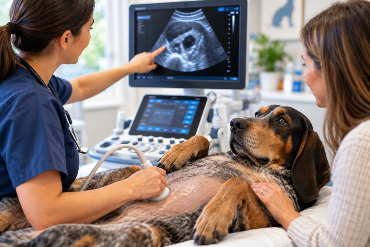
When a dog or cat needs an ultrasound or echocardiogram, many pet owners think of the examination as the moment when they will finally get an answer.
Sometimes that happens.
Other times, the scan answers one question while creating a more specific one.
A veterinary ultrasound may reveal a mass, abnormal organ, thickened intestinal wall, fluid accumulation, bladder stone, enlarged lymph node, or another unexpected finding. An echocardiogram may identify changes involving the heart valves, chamber size, wall thickness, blood flow, or pumping function.
Those findings can be extremely valuable, but an abnormal image does not always equal a final diagnosis.
Veterinary diagnostics often work as a sequence. The physical examination, medical history, laboratory testing, radiographs, ultrasound, echocardiogram, and other diagnostics each provide different pieces of information. The veterinary team then combines those pieces to determine what the finding most likely means and what should happen next.
That next step could involve additional blood work, fine needle aspiration, cytology, biopsy, CT imaging, surgery, medication, repeat imaging, or monitoring.
Understanding that process can make an ultrasound or echocardiogram result easier to understand and can help pet owners see why additional testing is sometimes recommended even after an advanced imaging study has already been performed.
An Abnormal Finding Is Not Always a Diagnosis
One of the most important concepts in veterinary imaging is the difference between finding an abnormality and identifying the disease causing it.
Ultrasound can provide detailed information about the appearance and structure of internal organs and tissues. It can identify changes in size, shape, architecture, fluid, masses, and other abnormalities.
However, many ultrasound findings are not specific to one disease.
For example, a mass may be visible on an ultrasound examination, but the appearance of that mass alone may not establish whether it is:
- Benign
- Malignant
- Inflammatory
- Infectious
- A different type of tissue abnormality
Similarly, thickening of an intestinal wall can be an important finding, but the image may not always establish the exact underlying cause.
This is why veterinary imaging is often described in terms of differential diagnoses.
A differential diagnosis is a list of conditions that could potentially explain the observed findings.
The imaging study helps narrow that list.
Additional testing may then help determine which possibility is most likely.
That distinction is important because it explains why a veterinarian may recommend another diagnostic procedure after an ultrasound that has already identified a problem.
What an Ultrasound Can Tell the Veterinary Team
Ultrasound is particularly useful because it produces dynamic, real-time images of soft tissues and internal organs.
The examination can provide information about structures such as the:
- Liver
- Gallbladder
- Spleen
- Kidneys
- Bladder
- Pancreas
- Intestinal tract
- Lymph nodes
- Other abdominal structures
Ultrasound can also identify findings such as fluid accumulation, masses, organ enlargement, intestinal abnormalities, urinary obstruction, and other structural changes.
But the value of the examination is not simply whether something looks abnormal.
The veterinary team also needs to consider where the abnormality is located, how extensive it appears to be, what structures surround it, and whether other abnormalities are present elsewhere.
That broader assessment can change what happens next.
For example, discovering one isolated abnormality may lead to a different treatment discussion than finding abnormalities affecting several organs.
The Difference Between a Finding, a Differential, and a Diagnosis
These three terms are useful for understanding veterinary imaging.
Finding
A finding is something abnormal that is observed during the examination.
Examples include:
- A mass
- Enlarged lymph nodes
- Thickened intestinal walls
- Fluid
- An enlarged organ
- Abnormal gallbladder contents
- Changes in kidney structure
Differential diagnosis
A differential diagnosis is a possible explanation for the finding.
There may be several possible explanations, particularly when an imaging appearance is not specific to one disease.
Confirmed diagnosis
A confirmed diagnosis is established when the available evidence is sufficient to identify the underlying disease or condition with an appropriate level of confidence.
Depending on the situation, confirmation may require laboratory testing, cytology, tissue biopsy, additional imaging, or other diagnostic information.
This progression is one reason an ultrasound should not always be judged by whether it immediately produces a single disease name.
A valuable ultrasound may instead take the diagnostic process from “something is wrong” to “we know where the problem is and what we need to investigate next.”
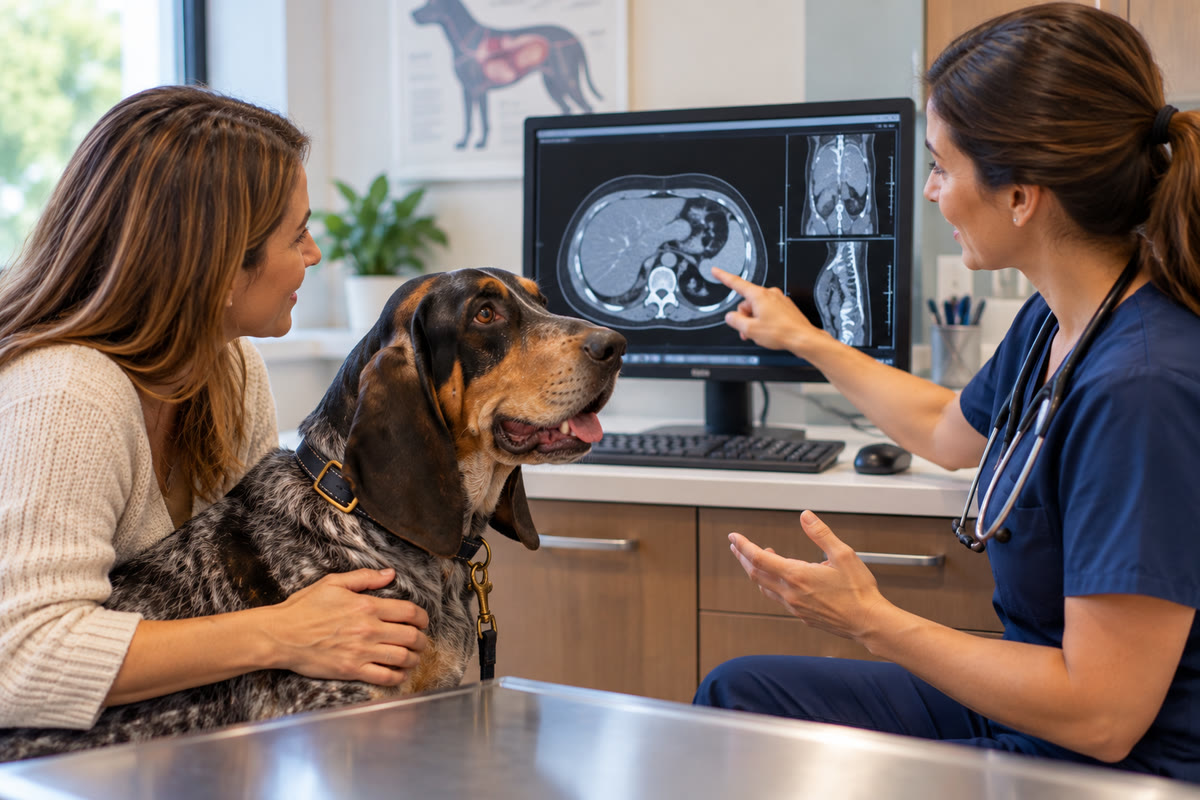
Why a Mass May Require Cytology or Biopsy
Finding a mass can be one of the most stressful outcomes for a pet owner.
However, a mass seen on ultrasound does not automatically mean cancer.
Ultrasound can show where a mass is located, how large it appears, its relationship to nearby structures, and certain characteristics of its appearance. Those details can help the veterinary team determine what possibilities need to be considered.
But imaging alone may not identify the exact cellular or tissue type.
That is where sampling can become important.
A veterinarian may recommend:
- Fine needle aspiration
- Cytology
- Biopsy
- Additional blood work
- CT imaging
- Follow-up imaging
The appropriate option depends on the location and characteristics of the abnormality, the pet's overall condition, and what information is needed to make the next medical decision.
In some cases, ultrasound can also help guide a needle toward a specific lesion.
This is an important advantage of ultrasound because the operator can visualize the target during the procedure rather than relying on an approximate location. Ultrasound-guided sampling can allow tissue or cell collection from lesions that may otherwise be difficult to target accurately.
The imaging examination therefore can become more than a diagnostic step. It can also help determine where additional diagnostic information should be obtained.
Why Ultrasound-Guided Sampling Can Be Valuable
Imagine that an ultrasound identifies an abnormal area inside a large organ such as the liver or kidney.
Knowing that the abnormality exists is useful.
But if the next question is what that abnormal tissue actually represents, the veterinary team may need cells or tissue for further evaluation.
Ultrasound can provide real-time visualization of the target while a sampling procedure is performed.
This can help the veterinarian identify the intended lesion and avoid simply sampling an unrelated area.
The broader principle is important:
Imaging can identify the target; laboratory or pathology testing can help identify the disease.
Those two types of information complement each other.
This is also why a recommendation for sampling after an ultrasound should not necessarily be interpreted as a failure of the imaging study. In many cases, the ultrasound successfully identified the exact area that requires further investigation.
When Ultrasound Changes the Surgical Conversation
Imaging can also influence whether surgery is considered and how a procedure may be approached.
Before surgery, veterinarians need to understand as much as possible about the location and extent of a problem.
Ultrasound may help identify:
- Which organs are involved
- Whether an abnormality appears localized
- Whether multiple areas are affected
- Whether fluid or bleeding is present
- Whether an obstruction is present
- Whether additional diagnostic information is needed
This information can help with surgical planning and with determining whether surgery is appropriate.
For example, an abdominal abnormality that appears localized may create a different treatment discussion from one that appears widespread.
Presurgical imaging can also provide information that may not be obvious during an initial physical examination.
The purpose is not simply to determine whether a pet has a problem. It is to gather enough information to make the next intervention as informed as possible.
Ultrasound Has Important Technical Limitations
Ultrasound is powerful, but it is not able to visualize everything equally well.
Sound waves interact differently with different tissues. Gas and bone can interfere with ultrasound transmission, which can make some structures difficult or impossible to evaluate completely. Intestinal gas, for example, can limit visualization of structures behind it.
Patient size, movement, anatomy, and the location of the abnormality can also affect the examination.
This creates an important diagnostic principle:
A structure that cannot be adequately visualized is not necessarily a normal structure.
Sometimes the limitation is simply that the ultrasound beam cannot obtain sufficient information from that particular area.
This is one reason veterinarians may recommend another imaging modality when ultrasound cannot answer the specific question being asked.
Depending on the case, that could include radiography, CT, MRI, or another diagnostic approach.
The goal is to choose the diagnostic method that provides the information still missing.
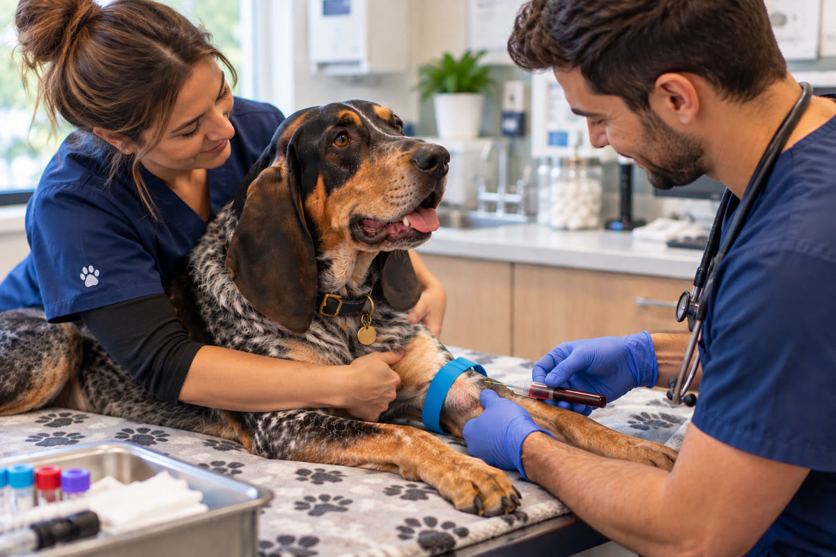
Why Ultrasound Findings Sometimes Do Not Match Blood Work Perfectly
Pet owners may also encounter situations where laboratory results and ultrasound findings do not appear to agree.
For example, blood work may strongly suggest disease while the ultrasound does not show an obvious corresponding abnormality.
The opposite can also happen.
An ultrasound may identify structural changes even when laboratory abnormalities are limited or absent.
This does not automatically mean one test is wrong.
Different diagnostic tests measure different aspects of disease.
Blood work provides information about physiological and biochemical changes.
Ultrasound provides information about anatomy and structure, along with real-time movement and other imaging characteristics.
Some diseases can be difficult to characterize through ultrasound alone, while other conditions may produce structural changes that are not fully reflected in routine laboratory testing.
Veterinary diagnostic imaging references specifically recognize that ultrasound findings and clinicopathologic findings do not always correspond perfectly in some diseases.
The veterinary team therefore looks at the combined evidence, rather than expecting every diagnostic test to produce identical results.
An Ultrasound Can Be Valuable Even When It Does Not Find the Original Suspected Problem
Another important but often overlooked benefit of diagnostic imaging is the ability to narrow the field of possibilities.
Suppose a veterinarian is concerned about an obstruction, internal bleeding, or another serious abdominal condition.
If ultrasound does not show evidence supporting that concern, that information can still influence the diagnostic plan.
The team can begin considering other explanations for the pet's symptoms.
In other words, diagnostic testing does not only identify what is present.
It can also help determine what is less likely.
This can prevent the veterinary team from continuing to pursue one explanation when the available evidence points somewhere else.
Unexpected Findings Can Change the Original Diagnostic Plan
Sometimes the most important finding is not the one the veterinary team expected to discover.
A pet may be referred for imaging because of elevated liver values, chronic vomiting, weight loss, urinary problems, or another concern.
During the examination, an unexpected abnormality may be identified.
Veterinary ultrasound can reveal findings such as:
- Tumors or masses
- Gallbladder disease
- Bladder stones
- Enlarged organs
- Fluid accumulation
- Hidden intestinal abnormalities
- Other structural changes
An unexpected finding can change the diagnostic priorities.
The veterinary team may need to determine whether the new abnormality is related to the original symptoms, whether it requires immediate attention, and whether additional testing is necessary.
This is one reason a systematic ultrasound examination can provide value beyond simply looking for one suspected condition.
Echocardiograms Answer Different Questions About the Heart
An echocardiogram is a specialized form of ultrasound used to evaluate the heart.
It provides real-time information about cardiac structures and their movement.
An echocardiogram can evaluate:
- Heart chamber dimensions
- Heart wall thickness
- Valve structure and movement
- Blood flow
- Pumping function
- Certain congenital abnormalities
- Pericardial fluid
- Some cardiac masses
- Other structural and functional abnormalities
More advanced echocardiographic techniques can also measure blood-flow velocities and provide quantitative information about cardiac function.
Doppler echocardiography is particularly useful because it evaluates the movement of blood through the heart and vessels. Abnormal flow patterns can help veterinarians evaluate conditions involving valves, narrowing, or other changes in cardiovascular blood flow.
This means an echocardiogram is not simply a picture of the heart.
It can provide information about how the heart is working.
Why an EKG and Echocardiogram Are Not the Same Test
Pet owners sometimes hear both “EKG” and “echocardiogram” and assume they evaluate the same problem.
They do not.
An electrocardiogram, or EKG/ECG, records the electrical activity of the heart.
It is particularly useful for identifying abnormalities in heart rhythm and electrical conduction.
An echocardiogram evaluates the heart's physical structures and function.
It can show chamber dimensions, wall thickness, valve motion, blood flow, and other structural characteristics.
That means one test may be useful when the concern is an arrhythmia, while another may be more appropriate when the concern involves valve disease, cardiomyopathy, chamber enlargement, or another structural problem.
Veterinary cardiovascular diagnosis often relies on multiple pieces of information, including history, physical examination, radiographs, ECG, laboratory testing, and echocardiography.
What an Abnormal Echocardiogram May Tell the Veterinary Team
An echocardiogram can help determine not only whether a cardiac abnormality exists, but also how significant certain changes may be.
For example, the examination can provide information about:
- Whether heart chambers are enlarged
- Whether heart walls are thickened
- Whether a valve is functioning abnormally
- Whether blood is moving through the heart at an abnormal velocity
- Whether heart muscle function is reduced
- Whether there are structural abnormalities
- Whether certain findings are consistent with particular forms of heart disease
This information can influence decisions about medication, monitoring, additional cardiac testing, or follow-up examinations.
Importantly, cardiac imaging is interpreted alongside the pet's clinical condition.
A measurement or image should not be considered in isolation from the patient's symptoms, physical examination, medical history, and other diagnostic results.
When the Best Next Step Is Monitoring
Not every abnormal finding requires immediate intervention.
Sometimes the most appropriate next step is to monitor the finding over time.
Repeat ultrasound may be recommended for:
- Gallbladder disease
- Chronic liver disease
- Tumor monitoring
- Benign-appearing nodules
- Response to treatment
- Other chronic conditions
Repeat echocardiography may also be useful for tracking cardiac disease as it changes over time.
The advantage of follow-up imaging is that the veterinary team can compare examinations.
Instead of asking only:
“What does this look like today?”
the team can also ask:
“Has this changed since the previous examination?”
That difference can be clinically meaningful.
An abnormality that remains stable may be managed differently from one that changes significantly over a short period.
Why Repeat Imaging Does Not Automatically Mean the Condition Is Worse
A recommendation for another ultrasound or echocardiogram can make owners worry that something has deteriorated.
But repeat imaging does not necessarily mean the disease has progressed.
Sometimes the purpose is surveillance.
A veterinarian may want to establish whether an abnormality is stable, changing slowly, responding to treatment, or developing characteristics that require a different approach.
For chronic disease, serial imaging can therefore become part of ongoing medical management rather than simply another diagnostic test.
This is particularly relevant when treatment decisions depend on whether an abnormality changes over time.
How Medical History Changes the Meaning of an Ultrasound
An ultrasound finding does not exist separately from the patient.
The veterinary team may consider:
- Age
- Species
- Breed
- Symptoms
- Duration of illness
- Physical examination findings
- Blood work
- Previous X-rays
- Previous imaging
- Current medications
- Relevant medical history
This information helps determine how an imaging finding should be interpreted.
For example, an abnormality discovered in a pet with chronic gastrointestinal symptoms may be interpreted differently from a similar finding in a pet with no gastrointestinal signs.
Likewise, an echocardiographic finding must be considered alongside the pet's clinical signs and other cardiovascular information.
The same image can have different clinical significance in different patients.
Why the Person Performing the Ultrasound Still Matters
Ultrasound is a real-time examination.
The operator moves the transducer, changes imaging angles, adjusts the examination based on what is seen, and decides when a particular structure needs closer evaluation.
This makes ultrasound different from a test in which a machine simply produces one standardized result.
An experienced examiner needs a strong understanding of normal anatomy and the ways disease can alter the appearance of tissues.
Ultrasound references also note that interpretation depends on recognizing normal echo patterns and understanding how abnormalities alter those patterns.
This matters because some abnormalities can be subtle.
Gas may interfere with visualization. A lesion may be small or located in a difficult area. Patient movement can affect image quality. Normal anatomy can vary between patients.
High-quality equipment is important, but obtaining useful diagnostic information also depends on appropriate technique and interpretation.
What Happens When the Ultrasound and Clinical Picture Do Not Match?
Sometimes the results create more questions instead of immediately resolving them.
A pet may have significant symptoms but limited ultrasound findings.
Another pet may have an abnormal ultrasound but relatively few outward signs.
When that happens, the veterinary team may reconsider the original diagnostic possibilities.
Additional testing may be recommended because the available evidence does not yet explain the complete clinical picture.
This is not necessarily a sign that the ultrasound was inconclusive or unhelpful.
It may mean that the disease process involves information that ultrasound cannot fully characterize.
The next diagnostic test is chosen based on the information that is still missing.
Questions to Ask After Your Pet's Ultrasound or Echocardiogram
When a pet owner receives an imaging result, the most useful question may not simply be, “Is it normal?”
Consider asking:
- What exactly did the ultrasound or echocardiogram show?
- Which organ or structure is affected?
- Is this an imaging finding or a confirmed diagnosis?
- What are the most likely explanations for this finding?
- Does the finding explain my pet's symptoms?
- Is additional testing necessary?
- Would cytology or biopsy provide useful information?
- Can the abnormality be sampled using ultrasound guidance?
- Is CT or another imaging method more appropriate?
- Does my pet need treatment now?
- Could this finding be monitored instead?
- When should repeat imaging be considered?
- What changes should I watch for at home?
These questions help turn an imaging report into a clearer understanding of the diagnostic plan.
The Goal Is Not Just to Find Something—It Is to Know What to Do With the Information
Veterinary ultrasounds and echos are valuable because they provide information that may not be available through a physical examination or routine laboratory testing alone.
But the most useful result is not necessarily the one that produces the longest list of abnormalities.
The real value comes from connecting the finding to the clinical question.
An ultrasound may:
Identify an abnormality → narrow the possible causes → guide sampling or additional testing → influence treatment → establish a baseline for monitoring.
An echocardiogram may:
Define cardiac structure → measure function and blood flow → assess the significance of disease → guide treatment and follow-up.
Sometimes imaging provides a clear answer.
Sometimes it tells the veterinary team what needs to be investigated next.
Sometimes it rules out a serious concern.
And sometimes a repeat examination provides information that a single snapshot could not.
That is why veterinary ultrasounds and echocardiograms are best understood as part of a larger diagnostic process. The scan provides another layer of information, and the veterinary team uses that information alongside the pet's history, examination, laboratory results, and other diagnostics to determine the most appropriate next step.