
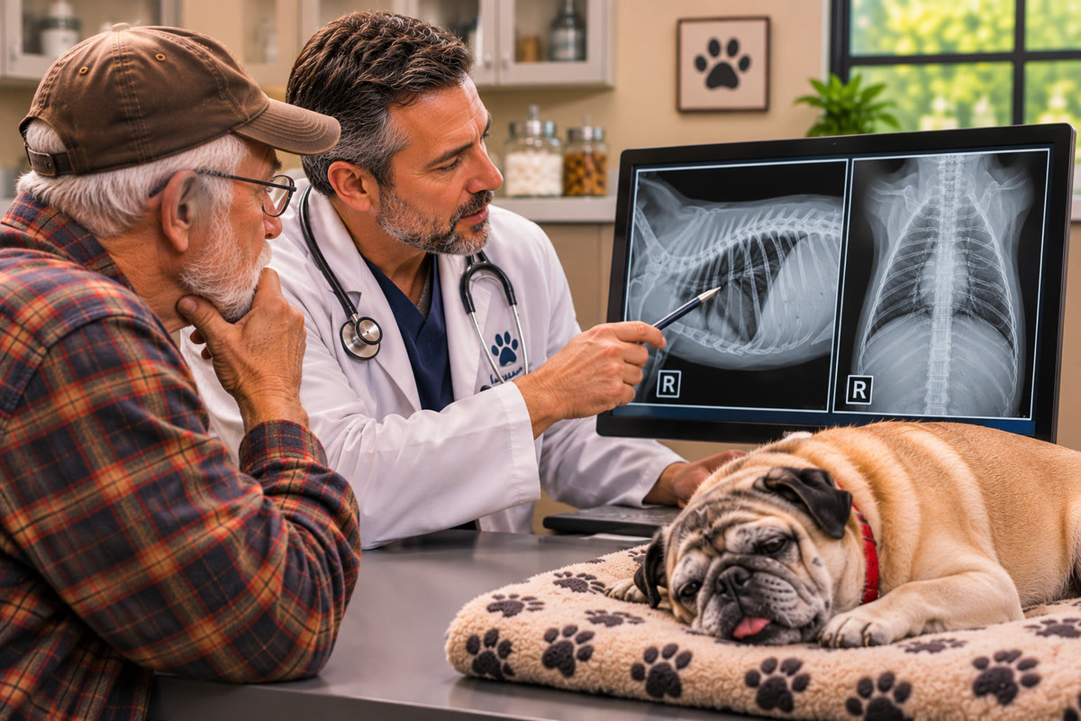
How Veterinary Ultrasounds and Echos Help Investigate Breathing Problems in Dogs and Cats
Breathing changes in dogs and cats can have several possible explanations. A pet may begin breathing faster than usual, struggle to get comfortable, lose interest in normal activities, or show increased effort when inhaling. These changes can involve the lungs, the space surrounding them, the heart, or the blood vessels responsible for circulation.
The difficulty is that respiratory symptoms often overlap. A dog with fluid accumulation in the lungs may breathe rapidly, but so might a dog with pneumonia or another pulmonary condition. A cat with heart muscle disease may develop respiratory distress, while another cat with similar signs may have a problem involving the pleural space.
This is why identifying the underlying cause requires more than recognizing a symptom.
Veterinary ultrasounds and echos provide different forms of information that can help investigate these conditions. Thoracic ultrasound examines accessible structures within the chest, including the pleural space and portions of the lung surface. An echocardiogram is a specialized cardiac ultrasound that evaluates the heart's chambers, valves, movement, and function.
These examinations are not interchangeable, and neither provides a complete picture of every respiratory disease. Their value comes from answering specific clinical questions and helping veterinarians determine which diagnostic steps are appropriate.

Why Respiratory Symptoms Require Careful Investigation
Normal breathing depends on several systems working together. Air must travel through the respiratory tract, the lungs must expand, oxygen must move into the bloodstream, and the heart must circulate oxygenated blood throughout the body.
A problem affecting any part of this process can change a pet's breathing pattern.
Some conditions primarily affect the lung tissue. Pneumonia, pulmonary edema, pulmonary hemorrhage, and certain tumors can change the amount of air, fluid, or tissue within the lungs.
Other conditions affect the pleural space, which is the narrow area between the lungs and the chest wall. Pleural effusion involves abnormal fluid accumulation in this space. Pneumothorax occurs when air enters the pleural space. Either condition can interfere with normal lung expansion.
Cardiovascular disease can also contribute to respiratory signs. For example, left-sided heart disease may increase pressure within the pulmonary circulation and lead to cardiogenic pulmonary edema. Other cardiovascular conditions may affect circulation or pressure within the pulmonary arteries.
Because these diseases can produce similar symptoms, veterinarians use the patient's history, physical examination, laboratory results, and imaging findings together. An ultrasound examination is most useful when it is selected to investigate a specific concern rather than treated as a stand-alone answer.
What Is Thoracic Ultrasound in Veterinary Medicine?
Thoracic ultrasound is an imaging examination that uses high-frequency sound waves to evaluate selected structures within the chest.
It is different from an abdominal ultrasound, which focuses on organs such as the liver, kidneys, bladder, and gastrointestinal tract. Thoracic ultrasound focuses on accessible areas of the chest wall, pleural space, lung surface, and other structures that can be visualized through appropriate acoustic windows.
There is an important technical limitation: normal lung tissue contains air, and air prevents ultrasound waves from traveling through the lungs in the same way they travel through fluid or soft tissue. As a result, ultrasound cannot provide a complete internal view of normal, air-filled lungs.
However, disease can change the relationship between air, fluid, and tissue near the lung surface. Those changes can create recognizable ultrasound findings.
Depending on the examination and the patient's condition, thoracic ultrasound may help identify:
- Fluid within the pleural space
- Findings associated with pneumothorax
- Irregularities along the pleural surface
- Increased vertical artifacts called B-lines
- Peripheral areas of lung consolidation
- Certain masses or abnormalities close to the chest wall
A 2025 clinical review of thoracic point-of-care ultrasound in small-animal emergency and critical care describes its applications in evaluating pleural space disorders and different pulmonary conditions. It also emphasizes that image interpretation depends on appropriate technique and the examiner's knowledge of thoracic anatomy and ultrasound artifacts.
This distinction is important: thoracic ultrasound does not replace every other form of chest imaging. It provides additional information about structures and abnormalities that can be evaluated using sound waves.
Understanding B-Lines and What They Can Reveal
One of the most discussed findings in veterinary lung ultrasound is the B-line.
B-lines appear as bright, vertical artifacts that originate from the pleural line and extend toward the bottom of the ultrasound screen. They move with lung sliding when the pleural surfaces move during breathing.
These artifacts are associated with changes in the peripheral lung's air-to-fluid or air-to-tissue relationship. They may become more numerous when there is increased fluid or tissue density in the lung's peripheral interstitial or alveolar regions.
Veterinarians may evaluate the number, distribution, and appearance of B-lines when investigating respiratory disease.
However, B-lines are not a diagnosis by themselves.
They can occur with cardiogenic pulmonary edema, but they may also be present with pneumonia, pulmonary contusions, hemorrhage, and other conditions affecting the lungs. Their significance depends on the patient's overall presentation and the distribution of the ultrasound findings.
A prospective study involving 200 dogs and cats with dyspnea evaluated lung ultrasound findings in patients with cardiogenic pulmonary edema, pneumonia, and lung neoplasia. The researchers found that a lung ultrasound score could help distinguish certain groups of patients, but its diagnostic accuracy varied by species and condition. The study reported fair-to-moderate accuracy for the conditions examined rather than a definitive ability to identify every cause of respiratory distress.
This is why a veterinarian should not interpret an increased number of B-lines as automatic proof of heart failure. The finding needs to be considered alongside cardiac imaging, radiographs, physical examination, and other available information.
How Thoracic Ultrasound Evaluates Pleural Effusion
Pleural effusion occurs when excessive fluid accumulates within the pleural space.
Normally, this space contains only a small amount of fluid that allows the pleural surfaces to move smoothly during breathing. When fluid accumulates, it can reduce the room available for the lungs to expand.
A pet with significant pleural effusion may show:
- Rapid or shallow breathing
- Increased respiratory effort
- Reduced activity
- Weakness
- Difficulty resting comfortably
- Open-mouth breathing in cats experiencing severe respiratory distress
Thoracic ultrasound can identify fluid between the lung and chest wall. It can also help the veterinary team assess where the fluid is located and whether there is an accessible area for sampling or drainage.
The appearance of pleural fluid may vary. Some fluid appears relatively dark and uniform on the ultrasound screen, while fluid containing cells, protein, debris, or fibrin may have a more complex appearance.
These visual differences can provide useful clues, but they do not reliably establish the fluid's exact composition or cause.
For example, an inflammatory effusion may contain cellular material, while blood or chyle can have different appearances depending on the sample and the clinical situation. There can also be overlap between fluid types.
When appropriate, thoracocentesis may be recommended. This procedure involves placing a needle or catheter into the pleural space to remove fluid. A sample can then be submitted for laboratory testing, including cytology and other analyses selected by the veterinarian.
Ultrasound can help guide the procedure, but fluid analysis is often necessary to investigate why the accumulation occurred.
Possible causes include inflammation, infection, trauma, cancer, and certain cardiovascular or systemic conditions.
Pneumothorax: When Air Accumulates Around the Lungs
Pleural effusion involves fluid in the pleural space. Pneumothorax involves air accumulating in that same space.
Normally, the negative pressure within the pleural cavity helps the lungs remain expanded against the chest wall. When air enters this space, the normal relationship between the lungs and chest wall can be disrupted. Depending on the amount of air and the underlying cause, part or all of a lung may collapse.
Pneumothorax can develop after trauma, certain medical procedures, or spontaneous leakage from damaged lung tissue.
Thoracic ultrasound may help identify findings associated with pneumothorax. One important feature is the assessment of lung sliding, which refers to the movement seen between the pleural surfaces during breathing.
When pneumothorax is present, lung sliding may be absent in the affected area because air separates the pleural surfaces. However, absent lung sliding is not specific to pneumothorax. Other factors, including severe lung disease, adhesions, and technical limitations, may also affect this finding.
This is why the examiner must assess multiple locations and interpret the result with the patient's condition and other imaging findings.
A 2021 study evaluating the Vet BLUE lung ultrasound protocol found that thoracic ultrasound could identify several types of thoracic abnormalities, but its performance varied by condition. The authors concluded that the protocol should not be used as the sole imaging method.
In a patient with suspected pneumothorax, the veterinarian may combine ultrasound findings with thoracic radiographs or other imaging when the patient's condition allows.
Lung Consolidation: When Air-Filled Tissue Changes Appearance
Healthy lung tissue contains air, which limits ultrasound visualization beyond the pleural surface. When a portion of the lung loses some of its normal air content and becomes denser, it may become visible as a tissue-like area on ultrasound.
This finding is called lung consolidation.
Consolidation can occur when air spaces become filled with fluid, inflammatory material, blood, or other substances. It may also develop when lung tissue becomes compressed or loses normal aeration.
Depending on the underlying condition, ultrasound may show different patterns. These can include:
- A tissue-like appearance within the affected area
- Irregularity along the pleural line
- Small areas of consolidation near the lung surface
- Air bronchograms, which appear as bright structures within consolidated tissue
- Associated B-lines or pleural fluid
Pneumonia is one possible cause of consolidation, but it is not the only one. Atelectasis, pulmonary hemorrhage, and other lung conditions may produce overlapping findings.
The movement of air bronchograms may provide additional information. Dynamic air bronchograms, which move with breathing, can support pneumonia in the appropriate clinical setting. However, no single ultrasound feature should replace the full diagnostic assessment.
A 2024 review of veterinary and human literature on lung ultrasound and subpleural consolidations notes that veterinary research in this area remains limited compared with human medicine. This is an important consideration when interpreting the examination: some findings are promising, but the evidence does not support treating every ultrasound pattern as a definitive diagnosis.
Additional testing may include chest radiographs, laboratory work, respiratory sampling, or other diagnostic procedures depending on the suspected disease.

When an Echocardiogram Is Needed
Thoracic ultrasound evaluates accessible structures within the chest, but it does not provide the same detailed assessment of the heart as a complete echocardiogram.
An echocardiogram is a specialized cardiac ultrasound that allows the examiner to evaluate the heart while it is beating.
It provides information about:
- Heart chamber dimensions
- Thickness of the heart walls
- Movement and appearance of the valves
- Cardiac contraction and relaxation
- Blood flow through selected structures
- Certain congenital abnormalities
- Findings associated with cardiovascular disease
An echocardiogram may be recommended when a veterinarian detects a heart murmur, suspects structural heart disease, or needs to investigate whether cardiovascular changes could be contributing to respiratory symptoms.
For example, left-sided heart disease can increase pressure within the pulmonary veins. In some patients, this can lead to cardiogenic pulmonary edema, where fluid accumulates within the lung tissue.
An echocardiogram can help identify relevant heart abnormalities, but it does not independently confirm every cause of pulmonary edema. The findings must be considered alongside the respiratory examination and other imaging.
This is especially important because not all lung fluid is caused by heart disease. Non-cardiogenic pulmonary edema and other pulmonary conditions can produce overlapping ultrasound findings.
Pulmonary Hypertension and the Role of Echocardiography
Pulmonary hypertension refers to increased pressure within the pulmonary circulation. It can develop in association with cardiac disease, respiratory disease, blood clots, parasitic disease, and other conditions.
Dogs with pulmonary hypertension may show signs such as exercise intolerance, increased breathing effort, respiratory distress, fainting, or signs of right-sided heart failure. However, these symptoms are not exclusive to pulmonary hypertension.
Echocardiography plays an important role in assessing the probability of pulmonary hypertension in dogs.
The ACVIM consensus statement on the diagnosis, classification, treatment, and monitoring of pulmonary hypertension in dogs recommends using echocardiography as part of a broader clinical assessment. The guidance emphasizes that echocardiography should be used to estimate the probability of pulmonary hypertension rather than treated as a definitive confirmation of the condition.
The examination may evaluate findings involving the right ventricle, pulmonary artery, right atrium, and blood-flow velocities measured using Doppler techniques.
These findings are interpreted together rather than relying on a single measurement. The consensus statement also highlights limitations in estimating pulmonary artery pressure through echocardiography, including potential variability and measurement imprecision.
This matters because an abnormal measurement does not automatically establish the severity or cause of pulmonary hypertension. The veterinarian must consider the patient's clinical signs, other cardiac findings, and results from additional diagnostic testing.
The consensus guidance applies specifically to dogs. Diagnostic decisions for cats require consideration of the available feline-specific evidence and the individual patient's condition.
Pericardial Effusion: Fluid Around the Heart
The heart is surrounded by a protective sac called the pericardium. A small amount of fluid normally exists between its layers.
Pericardial effusion occurs when an abnormal amount of fluid accumulates within this space.
When the pressure surrounding the heart becomes excessive, the heart may have difficulty filling with blood. This can lead to cardiac tamponade, a serious condition that interferes with normal circulation.
Affected pets may develop weakness, collapse, poor circulation, or other signs of cardiovascular compromise.
An echocardiogram is particularly useful for identifying fluid around the heart and assessing whether the accumulation is affecting cardiac filling.
The presence of pericardial fluid does not automatically mean that cardiac tamponade is occurring. The amount of fluid, the rate at which it accumulates, and the pressure it creates all influence its clinical significance.
When tamponade is present, urgent treatment may be required. In appropriate cases, pericardiocentesis may be performed to remove fluid from the pericardial space. Ultrasound guidance can help the veterinarian visualize the fluid and surrounding structures during the procedure.
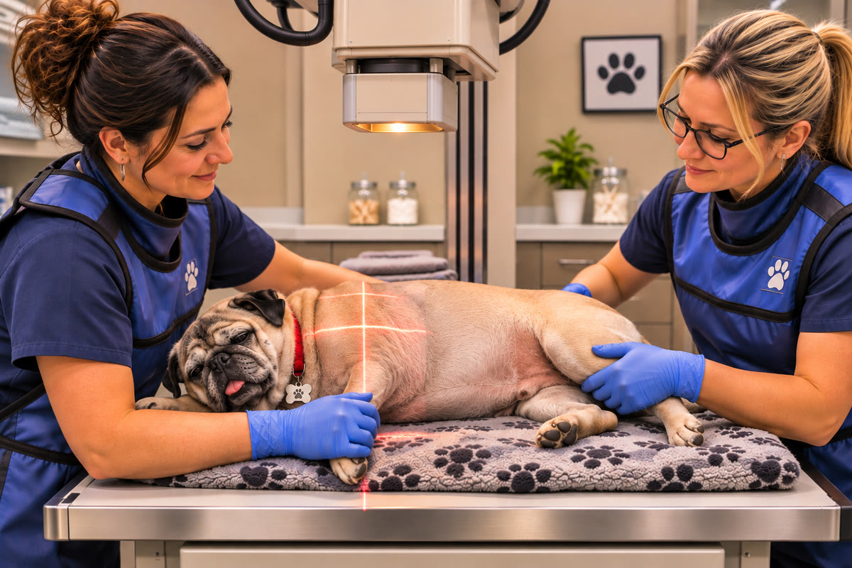
Why Veterinarians May Recommend Both Chest Imaging and an Echocardiogram
Chest radiographs, thoracic ultrasound, and echocardiography provide different information.
Thoracic radiographs offer a broader view of the chest. They can help evaluate lung patterns, heart size and shape, the position of major structures, and certain abnormalities involving the chest cavity.
Thoracic ultrasound provides real-time information about accessible lung surfaces, pleural fluid, and selected abnormalities near the chest wall.
Echocardiography focuses on the heart's internal structures and function.
A veterinarian may recommend more than one examination when the initial findings do not fully explain the patient's symptoms.
For example, a dog with rapid breathing may have a chest radiograph showing an abnormal lung pattern. Thoracic ultrasound may provide additional information about the peripheral lung and pleural space, while an echocardiogram may help determine whether structural heart disease or other cardiac changes are also present.
The decision depends on the patient's stability and the diagnostic question. A pet experiencing severe respiratory distress may not tolerate extensive handling or positioning. Stabilization and immediate treatment take priority over completing every possible imaging examination.
What Happens During a Thoracic Ultrasound or Echocardiogram?
The examination begins with a review of the patient's medical history, symptoms, previous imaging, and relevant laboratory results.
For thoracic ultrasound, the examiner evaluates selected areas of the chest using an ultrasound probe. The patient may be standing or resting in a comfortable position, depending on its condition and the examination protocol.
The examiner looks for findings such as pleural fluid, lung sliding, B-lines, pleural irregularities, and areas of consolidation.
An echocardiogram follows a more specialized approach. The examiner obtains standard views of the heart from different positions and evaluates cardiac chambers, valves, wall thickness, movement, and blood flow.
Many pets tolerate ultrasound examinations without sedation. However, the decision depends on the individual patient's comfort, anxiety, ability to remain still, and medical condition.
For pets with breathing difficulty, minimizing stress and unnecessary restraint is especially important. The veterinary team may adjust positioning or prioritize a limited examination based on the patient's stability.
Why Experience and Interpretation Matter in Veterinary Chest Imaging
Thoracic ultrasound requires a strong understanding of both anatomy and ultrasound physics. Air, bone, movement, and patient positioning can affect the images and limit which structures can be assessed.
The examiner must recognize normal pleural movement, identify artifacts, distinguish different patterns, and understand when a finding may have more than one explanation.
The same principle applies to echocardiography. Accurate assessment requires appropriate imaging views, consistent measurements, and interpretation of cardiac findings in the context of the patient's clinical condition.
Research also shows why experience and diagnostic judgment matter. Published studies of lung ultrasound in dogs and cats have reported useful applications, but diagnostic performance varies depending on the condition being investigated and the examination protocol used.
A 2021 study of the Vet BLUE protocol, for example, found that thoracic ultrasound could help identify lung and pleural abnormalities but should not be used as the only imaging method. Another prospective study found that B-line scoring could help distinguish certain causes of dyspnea, while its accuracy remained limited for other diagnostic comparisons.
These findings reinforce an important point: advanced imaging is most useful when the examiner understands both what the images show and what they cannot establish.
How Veterinary Diagnostic Centers Supports Diagnostic Evaluation
When a dog or cat develops breathing changes, identifying the underlying cause is an important part of determining the next steps in care.
Veterinary ultrasounds and echos can provide complementary information about the chest and cardiovascular system. Thoracic ultrasound may help evaluate pleural fluid, lung surface abnormalities, and selected pulmonary findings. Echocardiography provides a more detailed assessment of heart structure and function.
At Veterinary Diagnostic Centers, diagnostic imaging supports veterinarians who need additional information to investigate their patients' conditions. The appropriate examination depends on the referral question, the patient's clinical presentation, and the information needed to guide further evaluation.
For pet owners, the important thing is to seek veterinary attention when breathing changes occur rather than waiting for symptoms to become more severe. Rapid or labored breathing, open-mouth breathing in cats, collapse, or an inability to rest comfortably can indicate an urgent medical problem.
The right imaging approach helps the veterinary team investigate the structures involved, interpret findings in context, and determine whether additional testing or treatment is needed.