
When a dog or cat develops unexplained symptoms, abnormal blood work, or a new concern during a physical examination, the next diagnostic step is not always obvious.
A veterinarian may already have information from a physical examination, blood work, or X-rays, yet still need to understand what is happening inside the body. This is where advanced imaging can add information that other tests cannot provide on their own.
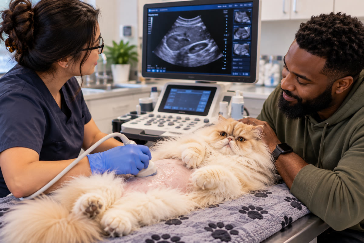
A veterinary ultrasound can allow the team to examine internal organs and soft tissues in real time. An echocardiogram, which is a specialized cardiac ultrasound, can provide detailed information about the structure and function of the heart.
These examinations are not necessarily replacements for other diagnostic tests.
They are pieces of a larger diagnostic process.
The value of ultrasounds and echos often comes from what they add to information that has already been collected—and what their findings help the veterinary team decide to do next.
Diagnosis Is Built From Multiple Pieces of Information
Veterinary medicine rarely depends on one finding in isolation.
A physical examination may identify a concern that needs further investigation. Blood work may reveal that an organ is not functioning normally. X-rays may show changes involving the chest or the overall size and shape of certain structures.
But these tests may not completely explain the underlying problem.
Ultrasound can provide another perspective by allowing the veterinary team to evaluate many organs and tissues directly and dynamically.
For example:
- Abnormal blood work may lead to an abdominal ultrasound.
- Persistent vomiting may prompt evaluation of the gastrointestinal tract and surrounding organs.
- Urinary symptoms may lead to imaging of the kidneys and bladder.
- A heart murmur may lead to an echocardiogram.
- An unexpected finding on another diagnostic test may create a new reason to examine a specific organ more closely.
The important point is that the imaging examination is connected to the clinical question.
The veterinarian is not simply looking for “something abnormal.” The goal is to gather information that helps explain the patient's symptoms, clarify an existing concern, or determine what should happen next.

What Blood Work Can Tell You Before an Ultrasound
Blood work can provide valuable information about how a pet's body is functioning.
It may identify changes involving organs, blood cells, or other body systems. Sometimes these abnormalities are discovered even when a pet does not appear seriously ill at home.
However, laboratory results do not always identify the structural reason for an abnormality.
Consider a pet with elevated liver values.
The blood work can tell the veterinarian that further investigation may be warranted. It cannot necessarily show whether the change is associated with the liver itself, the gallbladder, another organ, inflammation, or another disease process.
An abdominal ultrasound can provide additional information by allowing the veterinary team to examine the liver, gallbladder, and surrounding structures.
This creates a useful distinction:
Laboratory testing can identify that something is abnormal. Imaging can help investigate what may be happening structurally.
The two are not interchangeable.
They answer different questions.
Why an Ultrasound May Be Recommended After an X-Ray
Pet owners sometimes wonder why another imaging test is necessary when their dog or cat has already had X-rays.
The answer is that X-rays and ultrasound provide different types of information.
X-rays can be useful for evaluating bones, the chest, overall organ size and shape, and certain other abnormalities.
Ultrasound provides a different view of many soft tissues and internal organs. It also allows structures to be evaluated in real time while the examination is being performed.
For a pet with gastrointestinal symptoms, for example, X-rays may provide useful information about the abdomen and may help identify certain abnormalities. Ultrasound can provide additional detail about organs and soft tissues that require closer examination.
The veterinary team may therefore use both tests when the information from one examination does not completely answer the clinical question.
The goal is not to perform as many tests as possible.
It is to obtain the type of information needed to investigate the problem appropriately.
What a Veterinary Ultrasound Can Actually Show
Abdominal ultrasound can be used to examine structures such as:
- Liver
- Gallbladder
- Kidneys
- Bladder
- Pancreas
- Intestinal tract
- Spleen
- Lymph nodes
- Other abdominal structures
Depending on the patient and the reason for the examination, ultrasound may identify findings such as:
- Masses or tumors
- Inflammation
- Fluid accumulation
- Enlarged organs
- Bladder stones
- Urinary obstruction
- Intestinal abnormalities
- Other structural changes
But identifying an abnormality is only one part of the process.
The more important question is often:
What does this finding mean for this particular patient?
That requires the imaging findings to be considered alongside symptoms, medical history, physical examination findings, laboratory results, and previous diagnostics.
Ultrasound Is a Dynamic Examination
One of the distinguishing features of veterinary ultrasound is that the examination takes place in real time.
The person performing the scan can change the angle of the probe, evaluate a structure from different directions, examine an area more closely, and adjust the examination when something unexpected appears.
This matters because an abnormality may not be located exactly where the veterinary team initially expected it to be.
A patient may be referred because of abnormal liver values, for example, but the examination can reveal an additional concern involving another abdominal structure.
The scan can then receive additional attention in that area.
This is one reason ultrasound is highly operator-dependent.
The examination involves ongoing technical decisions rather than simply collecting a fixed set of images.
Why Operator Experience Matters
Ultrasound equipment is important, but the machine itself does not interpret the patient.
The person performing the examination must understand normal anatomy, recognize abnormalities, adjust the examination when necessary, and determine which areas require closer evaluation.
This becomes particularly important when visualization is difficult.
Factors such as intestinal gas, patient movement, body size, anatomy, or a small focal abnormality can make certain structures more challenging to evaluate.
An experienced sonographer can adapt the examination to these circumstances.
That may involve changing imaging angles, spending additional time on a suspicious structure, tracking difficult intestinal structures, or focusing on areas that are particularly relevant to the patient's symptoms.
The quality of the equipment matters.
So does the skill and experience behind the examination.
What Happens When Ultrasound Finds Something Unexpected?
An ultrasound does not always confirm the problem that originally prompted the referral.
Sometimes it identifies something that was not suspected beforehand.
VDC's diagnostic materials describe findings such as tumors, gallbladder disease, bladder stones, enlarged organs, fluid accumulation, and hidden intestinal disease being identified during ultrasound examinations.
This can change the direction of the diagnostic workup.
For example, a pet may be referred because of abnormal liver values. The initial concern may be focused on the liver, but the examination could reveal an abnormality involving another abdominal organ.
That new finding may require additional investigation.
This is one of the practical advantages of a complete diagnostic imaging examination: it can provide information that changes the original understanding of the case.
An Abnormal Ultrasound Does Not Always Mean a Final Diagnosis
One of the most important limitations for pet owners to understand is that ultrasound does not always identify the exact disease responsible for an abnormal finding.
A scan may reveal a mass.
It may show abnormal lymph nodes.
It may identify thickened intestinal tissue, fluid, or an abnormal organ.
But imaging alone may not always determine:
- Whether a tumor is cancerous
- The exact type of disease
- The complete microscopic nature of an abnormality
This is why an abnormal ultrasound may be followed by additional testing.
Depending on the findings, the veterinary team may recommend:
- Fine needle aspiration
- Cytology
- Biopsy
- Additional blood work
- CT imaging
- Repeat ultrasound
- Other diagnostic procedures
The important distinction is that the ultrasound has still provided valuable information.
It may have identified where the problem is, shown what it looks like, and helped determine which question needs to be answered next.
From Imaging Finding to Next Diagnostic Step
The relationship between an ultrasound finding and the next diagnostic step is an important part of the overall workup.
Consider a few examples.
A Mass Is Identified
A mass may be visible on ultrasound, but its appearance may not establish exactly what type of tissue it contains.
The next step may involve sampling, cytology, or biopsy.
An Abnormality Appears Localized
If a problem appears confined to a particular organ or region, the veterinary team may have a clearer idea of where to focus additional diagnostics or treatment planning.
Multiple Structures Are Affected
If abnormalities appear in several areas, the diagnostic approach may need to consider whether there is a broader disease process.
A Serious Problem Is Ruled Out
Sometimes the value of imaging is not finding the suspected disease.
If a major concern is not identified, that information can help redirect the diagnostic investigation.
This is why the usefulness of ultrasound should not be measured only by whether it produces a single definitive diagnosis.
Sometimes the most important result is knowing what the veterinary team should investigate next.
How Ultrasound Findings Can Affect Treatment Decisions
Diagnostic imaging is also used to help guide treatment planning.
Ultrasound may help veterinarians evaluate:
- Which organs are involved
- Whether disease appears localized or widespread
- Whether additional testing is needed
- Whether surgery may need to be considered
- Whether monitoring is appropriate
- How urgent further care may be
For example, identifying internal bleeding, a ruptured organ, or an obstruction can create a very different treatment situation than identifying a finding that can be monitored.
The imaging result is not considered by itself.
It is interpreted alongside the patient's symptoms, physical examination, laboratory results, and overall condition.
The purpose is to give the veterinary team more information for making an informed medical decision.
Why Ultrasound Can Be Important in Emergency Cases
In emergency situations, the diagnostic question can be particularly urgent.
Ultrasound may help identify problems such as:
- Internal bleeding
- Ruptured organs
- Urinary obstruction
- Gastrointestinal perforation
- Foreign body obstruction
- Gallbladder rupture
- Other significant abdominal abnormalities
When these types of findings are suspected, the information obtained from imaging may help determine whether the patient needs urgent intervention, additional diagnostics, surgery, or medical management.
The importance of the scan comes from how quickly the information can change the understanding of the patient's condition.
Urinary Problems Are One Example of Targeted Ultrasound
Urinary symptoms can sometimes provide a clear reason for imaging.
Signs such as:
- Straining to urinate
- Blood in the urine
- Frequent urination
- Urinary accidents
- Suspected obstruction
may lead a veterinarian to investigate the urinary system more closely.
Ultrasound can help evaluate the bladder and kidneys and may identify findings such as bladder stones, urinary obstruction, masses, kidney abnormalities, or bladder inflammation.
In a patient with suspected urinary obstruction, identifying the problem quickly can be particularly important because an obstruction may require immediate treatment.
This illustrates how the value of ultrasound depends on the clinical question being asked.
When an Echocardiogram Is the More Appropriate Ultrasound
Not every ultrasound examines the abdomen.
An echocardiogram is a specialized cardiac ultrasound used to evaluate the heart's structure and function in real time.
Veterinary echocardiography can evaluate:
- Heart valves
- Heart wall thickness
- Chamber size
- Blood flow
- Pumping function
- Pulmonary pressures
- Heart muscle changes
An echocardiogram may be recommended when a veterinarian identifies a heart murmur, arrhythmia, exercise intolerance, coughing, increased respiratory effort, weakness, collapse, or another cardiovascular concern.
The purpose is to obtain information about the heart that cannot be fully provided by a general physical examination.
Echocardiogram vs. EKG: Why Both May Be Used
An EKG and an echocardiogram are sometimes confused because both are associated with cardiac testing.
They do not evaluate the same thing.
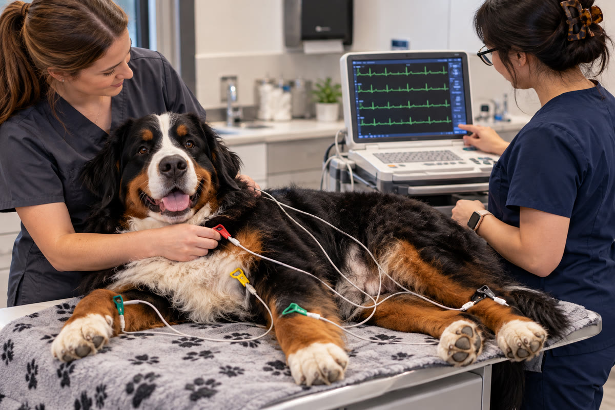
An EKG evaluates the heart's electrical activity and rhythm.
An echocardiogram evaluates the heart's physical structures and function.
This means a pet may need one test, the other, or both.
For example, an abnormal rhythm may make evaluation of electrical activity important, while a heart murmur may prompt an echocardiogram to examine valves, chambers, heart walls, and blood flow.
Using different cardiac tests does not necessarily mean that the previous test was insufficient.
It may simply mean that the veterinary team is asking a different clinical question.
What an Echocardiogram Can Add to a Heart Evaluation
A heart murmur is a finding, not a complete diagnosis.
An echocardiogram can help determine which cardiac structures are involved and provide information about how the heart is functioning.
This can help the veterinary team understand whether a cardiac condition appears mild or more advanced, determine which structures are affected, and decide whether medication or ongoing monitoring may be appropriate.
Cardiac disease can also differ between dogs and cats.
Degenerative valve disease is common in dogs, particularly involving the mitral valve, while cats more commonly experience cardiomyopathies involving the heart muscle.
Because different cardiac conditions affect the heart in different ways, detailed imaging can provide information that helps distinguish the type of problem being investigated.
Why Your Pet's History Changes How Imaging Findings Are Interpreted
An ultrasound image does not exist independently of the patient.
Two pets can have similar-looking abnormalities but very different medical histories.
One may have weight loss and abnormal blood work.
Another may have no obvious symptoms but an incidental finding discovered during an examination.
The context can influence what the veterinary team considers important and what additional testing may be appropriate.
That is why medical history, symptoms, physical examination findings, blood work, X-rays, and previous diagnostics remain relevant throughout the imaging process.
The goal is not simply to identify an abnormal image.
It is to understand the finding in the context of the patient.
When an Ultrasound Leads to Sampling or Biopsy
One of the clearest examples of ultrasound guiding the next diagnostic step is when an abnormal structure needs tissue or cell evaluation.
Suppose an ultrasound identifies a mass.
The scan may show its location and physical characteristics, but it may not establish whether the mass is benign, malignant, inflammatory, or another type of abnormal tissue.
A fine needle aspiration, cytology, or biopsy may therefore be recommended.
This creates a sequence:
Imaging identifies the abnormality → additional testing characterizes the abnormality → the veterinary team uses the combined information to guide care.
The tests are complementary.
The ultrasound does not need to answer every question for it to be diagnostically valuable.
Repeat Ultrasound Can Answer a Different Question
An initial ultrasound asks, in effect, “What is happening now?”
A follow-up examination may ask, “What has changed since the last examination?”
That distinction can be important for pets with conditions that require monitoring.
Repeat imaging may be recommended for:
- Gallbladder disease
- Chronic liver disease
- Tumor or mass monitoring
- Benign nodules
- Cardiac disease
- Response to treatment
- Other chronic conditions
A follow-up scan can help determine whether a finding has remained stable, changed over time, or responded to treatment.
Echocardiography may also require periodic reassessment for pets with cardiac disease because the condition may change over time.
The timing of repeat imaging depends on the patient's condition and the veterinary team's recommendations.
What Pet Owners Can Expect Before an Ultrasound
Preparation depends on the type of examination and the individual patient.
For an abdominal ultrasound, the veterinary team may review:
- Current symptoms
- Medical history
- Blood work
- Previous imaging
- Other diagnostic results
A small area of fur may be shaved to improve contact between the probe and the skin, and ultrasound gel is applied during the examination.
Many pets can undergo ultrasound without sedation.
However, sedation may sometimes be recommended when a pet is highly anxious, painful, unable to remain still safely, or needs another procedure at the same time.
The examination itself is non-invasive and generally well tolerated.
Your veterinary team will provide specific preparation instructions, including whether fasting is appropriate for your pet.
What Pet Owners Should Ask About the Results
When an ultrasound or echocardiogram is recommended, understanding the purpose of the test can make the process easier to follow.
Useful questions include:
What specific question are we trying to answer?
This helps clarify why the imaging has been recommended.
What will this test show that the previous tests did not?
This can help explain why another diagnostic method is necessary.
If something abnormal is found, what could the next step be?
Depending on the finding, the next step could involve sampling, biopsy, additional blood work, CT, treatment, or monitoring.
Does the finding require immediate action?
Some findings may require urgent attention, while others may be appropriate for further investigation or monitoring.
Could my pet need repeat imaging?
Some conditions are followed over time, particularly chronic disease, masses, gallbladder problems, and cardiac disease.
Will the ultrasound provide a definitive diagnosis?
Sometimes it can. In other cases, it narrows the possibilities and identifies what needs to be investigated next.