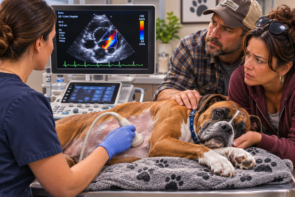
When a veterinarian recommends an ultrasound, many pet owners picture a black-and-white image of internal organs on a monitor. However, veterinary ultrasound can reveal more than the size and appearance of the structures being examined. Specialized techniques can also help evaluate how blood moves through the heart and blood vessels.
This is where Doppler imaging becomes important.
Doppler ultrasound provides information about the direction, pattern, and velocity of blood flow. It can help veterinarians investigate abnormal circulation, evaluate heart valves, assess blood movement through cardiac chambers, and examine selected abdominal blood vessels.
These measurements provide information that conventional grayscale ultrasound cannot show by itself. They are particularly valuable when a veterinarian needs to understand not only what a structure looks like, but also how blood moves through it.
Doppler techniques are used during abdominal ultrasound examinations and echocardiograms. Although they rely on the same underlying principle, their clinical applications differ depending on the structures being evaluated.
What Is Doppler Ultrasound in Veterinary Medicine?
Doppler ultrasound is an imaging technique that uses changes in the frequency of returning sound waves to estimate movement. In veterinary medicine, it is commonly used to evaluate blood flowing through vessels and within the heart.
The ultrasound machine sends sound waves into the body. When these waves encounter moving red blood cells, the reflected sound changes in frequency depending on the movement of the blood relative to the ultrasound probe. The machine processes these changes to provide information about blood-flow direction and velocity.
This differs from conventional grayscale ultrasound, which primarily displays the appearance of tissues and anatomical structures.
For example, a grayscale image may show a heart valve and the surrounding chambers. Doppler imaging can add information about whether blood is moving normally through the valve or whether an abnormal flow pattern is present. Similarly, an abdominal ultrasound may identify a blood vessel, while Doppler helps assess whether blood is flowing through it and whether the observed pattern raises concern.
The American College of Veterinary Radiology (ACVR) and European College of Veterinary Diagnostic Imaging (ECVDI) abdominal ultrasound consensus statement identifies Doppler as an important technique for evaluating vascular patency, vascular malformations, and tissue perfusion when clinically indicated.
This means Doppler is not simply an additional color display. It is a diagnostic tool used to investigate specific questions about circulation.
The Three Main Types of Doppler Imaging
Veterinary ultrasound examinations may use different Doppler modes depending on the clinical question, the location of the structure, and the type of blood-flow information required.
1. Color Doppler
Color Doppler displays blood flow as color information overlaid on the grayscale ultrasound image. It helps the examiner visualize the direction and distribution of blood flow relative to the ultrasound probe. It can also reveal flow patterns that appear turbulent or abnormal.
During an echocardiogram, color Doppler may help identify blood flowing backward through a valve, a finding known as regurgitation. It can also help evaluate abnormal flow through the heart's outflow pathways.
In abdominal imaging, color Doppler may help assess blood vessels and the vascular characteristics of selected lesions.
The colors do not represent a universal distinction between oxygen-rich and oxygen-poor blood. Instead, they indicate the direction of movement relative to the probe according to the machine's color map. The display is also affected by the settings selected during the examination, so color alone does not establish the severity of an abnormality.
2. Spectral Doppler
Spectral Doppler provides a waveform representing blood-flow velocity over time. Rather than displaying flow only as a color overlay, it allows the examiner to evaluate how fast blood is moving at a selected location along the ultrasound beam.
This can be useful when assessing blood flow across heart valves, through the heart's outflow tracts, or within certain abdominal vessels.
The waveform provides information about changes in velocity during different parts of the cardiac cycle. Its shape and pattern can also help characterize circulation within a vessel. The examiner interprets these measurements in relation to the anatomy and the clinical question being investigated.
3. Power Doppler
Power Doppler displays the strength of the Doppler signal rather than primarily emphasizing the direction and velocity of blood flow.
It can be useful for detecting relatively slow blood flow in certain situations and evaluating vascularity in selected tissues or lesions. However, it does not provide the same directional information as color Doppler or the velocity measurements available through spectral Doppler.
Each technique has a specific role. The choice depends on what the veterinary team needs to investigate.

How Doppler Echocardiography Evaluates the Heart
An echocardiogram is a specialized ultrasound examination of the heart. It evaluates cardiac structures and function in real time, including the chambers, valves, heart walls, and movement of blood through the heart.
Doppler echocardiography adds information about the blood moving through these structures. This is important because some heart abnormalities cannot be fully understood by examining the heart's shape alone.
Evaluating Heart Valve Function
Heart valves help direct blood through the heart in an organized sequence. When a valve does not close properly, some blood may flow backward instead of continuing in the intended direction. This is called valvular regurgitation.
Color Doppler can help visualize the direction and distribution of the abnormal flow. Spectral Doppler can provide additional measurements of blood-flow velocity.
For example, a veterinarian evaluating a dog with suspected myxomatous mitral valve disease may examine the mitral valve and assess whether blood is flowing backward into the left atrium during contraction.
Myxomatous mitral valve disease is a common acquired heart condition in dogs, particularly older small-breed dogs. As the valve changes over time, it may become thickened or fail to close properly. This allows blood to leak backward and can gradually contribute to enlargement of the left atrium and left ventricle.
The presence of a regurgitant jet is only one part of the evaluation. Its appearance and measured characteristics must be considered alongside valve structure, chamber dimensions, and the patient's clinical findings.
The American College of Veterinary Internal Medicine (ACVIM) consensus guidelines for myxomatous mitral valve disease recommend echocardiography performed by an experienced operator to identify the cause of a murmur, evaluate cardiac chamber enlargement, and investigate relevant complications.
This distinction matters because identifying valve leakage does not automatically establish how advanced the disease is. The veterinarian also needs to determine whether the heart has undergone measurable structural changes.
Why Chamber Measurements Matter Alongside Blood Flow
When a heart valve leaks, the receiving chamber may be exposed to additional blood volume. Over time, this can contribute to enlargement of the chamber.
An echocardiogram therefore evaluates more than the presence of regurgitation. Measurements of the left atrium and left ventricle help the veterinarian assess whether the heart is remodeling in response to the abnormal circulation.
In dogs with myxomatous mitral valve disease, ACVIM guidelines include measurements such as the left atrium-to-aorta ratio and body-weight-normalized left ventricular internal diameter when assessing clinically important cardiac enlargement.
These measurements help distinguish dogs with valve disease but limited cardiac remodeling from those with more substantial enlargement. Doppler helps characterize abnormal blood movement, while structural measurements show how the heart is responding to that abnormality.
Investigating Abnormal Flow Through the Heart
Doppler echocardiography can also help identify abnormal flow through the heart's outflow pathways.
When blood passes through a narrowed or obstructed area, its velocity may increase. Doppler measurements can help estimate the pressure difference across certain structures when the examination is technically appropriate.
This information can be important when evaluating suspected valve narrowing or obstruction within the heart. For example, a veterinarian may investigate increased blood-flow velocity through the aortic or pulmonary outflow tract when the physical examination or other findings raise concern.
The velocity measurement is not interpreted alone. The examiner also considers the structure involved, the imaging views, and whether the observed pattern is consistent with the suspected condition.
How Doppler Helps Investigate Heart Disease in Cats
Cats can develop heart muscle conditions that change the thickness, shape, and function of the heart. Hypertrophic cardiomyopathy is one example. It involves abnormal thickening of the heart muscle, particularly the left ventricular wall.
A standard echocardiogram allows the veterinarian to measure the heart walls and chambers. Doppler techniques provide additional information about blood movement through the heart.
In some cats, thickening of the heart muscle can contribute to dynamic left ventricular outflow tract obstruction. This occurs when the pathway through which blood leaves the left ventricle becomes narrowed during part of the cardiac cycle.
One mechanism associated with this obstruction is systolic anterior motion of the mitral valve. During contraction, part of the mitral valve moves toward the septum and can interfere with blood leaving the left ventricle.
The ACVIM consensus guidelines for feline cardiomyopathies recommend evaluating for dynamic left ventricular outflow tract obstruction using a combination of two-dimensional, M-mode, and Doppler echocardiography.
Color Doppler can help identify characteristic flow patterns associated with obstruction and mitral regurgitation. Spectral Doppler can estimate the peak pressure gradient across the affected area when appropriate views and measurements are obtained.
The guidelines also discuss assessing diastolic function using spectral Doppler and tissue Doppler imaging. Diastolic function describes how the heart relaxes and fills between contractions, which is particularly relevant in cats with cardiomyopathy.
Another important consideration is blood stasis within an enlarged left atrium. In some cats with significant cardiac disease, reduced atrial function can contribute to slow-moving blood and the formation of spontaneous echocardiographic contrast, sometimes described as a smoke-like appearance on ultrasound.
This finding can be associated with an increased risk of blood clot formation. In cats with left atrial enlargement, the consensus guidelines recommend evaluating the left atrial appendage for spontaneous echocardiographic contrast or thrombus and assessing blood-flow velocities when appropriate.
These findings demonstrate why a comprehensive echocardiogram involves more than measuring heart wall thickness. It may also assess blood movement, filling patterns, and signs of impaired circulation that can affect a cat's risk.
Doppler Ultrasound in Abdominal Imaging
Doppler imaging is not limited to the heart. It can also be used during abdominal ultrasound when a veterinarian needs to evaluate blood vessels, circulation, or the vascular characteristics of an abnormality.
Some abdominal conditions involve changes in the way blood moves through the liver or other organs. In these situations, grayscale imaging may show the anatomy, while Doppler provides additional information about circulation.
The ACVR and ECVDI consensus statement recommends using color, power, or spectral Doppler when there is a clinical question involving vascular patency, vascular malformations, or perfusion characteristics.
Evaluating the Portal Vein
The portal vein carries blood from the digestive organs and spleen toward the liver. This circulation is important because the liver processes substances absorbed from the gastrointestinal tract before blood returns to the general circulation.
Doppler ultrasound can help assess portal blood flow, including its direction, velocity, and waveform characteristics.
One condition that may require this evaluation is a portosystemic shunt. This is an abnormal connection that allows some blood from the portal circulation to bypass the liver.
Portosystemic shunts may be congenital or acquired. Congenital shunts are present from birth, while acquired shunts can develop in association with conditions that alter pressure within the portal circulation.
Depending on the location and type of shunt, Doppler may help identify unusual vessel connections or abnormal portal blood-flow patterns.
For example, blood normally travels toward the liver through the portal vein. In certain acquired shunting conditions associated with portal hypertension, the flow direction may become reversed, a pattern called hepatofugal flow. This finding can raise concern when interpreted alongside other clinical and imaging results.
However, a single flow measurement does not establish the presence or type of shunt.
Why Portosystemic Shunts Matter
When blood bypasses the liver, substances that would normally be processed by the organ may remain in circulation.
Some affected pets develop signs such as poor growth, gastrointestinal problems, unusual behavior, or neurological changes. The severity and timing of symptoms can vary.
Doppler ultrasound can contribute to the investigation by assessing the portal circulation and looking for abnormal vascular pathways. However, not every shunt can be completely characterized with ultrasound. Some vascular structures are difficult to visualize because of their location, size, or surrounding anatomy.
A review of imaging for portosystemic shunts in dogs and cats describes ultrasound as an important part of the diagnostic workup while also discussing the role of contrast-enhanced CT in defining more complex vascular anatomy.
CT angiography may be recommended when a more detailed map of the blood vessels is needed, particularly for treatment planning.
Assessing Blood Flow Around Abdominal Lesions
Doppler can also be useful when evaluating the blood supply to selected abdominal organs or investigating the vascular characteristics of a mass.
For example, a lesion may look different from the surrounding tissue on grayscale ultrasound. Doppler can help determine whether detectable blood flow is present within or around the area.
This information may contribute to the overall interpretation, but vascularity does not establish whether a lesion is benign or malignant.
Some tumors have substantial blood supply, while other lesions may show little detectable flow. The absence of a visible Doppler signal does not necessarily mean that a structure has no blood supply. Slow flow, technical settings, and the size of the vessels can affect what the machine detects.
The finding must be considered alongside the lesion's appearance, location, and the pet's clinical history. Additional testing, including sampling or other imaging, may be needed to determine what the abnormality represents.
Why Doppler Findings Need to Be Interpreted Carefully
Doppler imaging provides useful measurements, but those measurements are affected by several technical and patient-related factors.
The Angle of the Ultrasound Beam
The accuracy of Doppler velocity measurements depends on the angle between the ultrasound beam and the direction of blood flow.
When the beam is not aligned appropriately, the measured velocity may be underestimated. This is particularly important during echocardiography, where the examiner must obtain suitable views of moving blood through valves and outflow tracts.
A technically limited measurement should not be treated as a definitive representation of the patient's actual blood-flow velocity.
Machine Settings and Signal Quality
Doppler settings influence what the machine displays. For example, the velocity scale must be appropriate for the type of vessel or flow being evaluated.
If settings are not optimized, the image may show aliasing. This occurs when the displayed color or waveform cannot accurately represent the measured velocity within the selected scale.
Other factors, including gain, sample location, patient movement, and surrounding tissue, can also affect the image. These technical considerations explain why Doppler results require a trained examiner who understands how to obtain and interpret the signals.
Heart Rate and Patient Condition
Blood-flow measurements are influenced by the patient's cardiovascular state. Heart rate, rhythm, hydration, blood pressure, breathing, and other physiological factors can affect measurements or the way they are interpreted.
For example, a measurement obtained while a pet is highly stressed may differ from one obtained when the patient is calm. The veterinarian considers the patient's condition during the examination and interprets the results within that context.
Doppler Ultrasound Is Not the Same as an EKG
Pet owners sometimes hear the terms echocardiogram and EKG used together and assume they refer to the same test. They are different diagnostic procedures.
An electrocardiogram, commonly called an EKG or ECG, records the electrical activity of the heart. It helps evaluate heart rhythm and identify certain electrical abnormalities.
An echocardiogram uses ultrasound to examine the heart's physical structures and movement. Doppler echocardiography adds information about blood flow through the heart.
| Diagnostic test | What it evaluates | What it can help investigate |
|---|---|---|
| EKG/ECG | What it evaluates: Electrical activity of the heart | What it can help investigate: Arrhythmias and rhythm abnormalities |
| Standard echocardiogram | What it evaluates: Heart structure and movement | What it can help investigate: Chamber size, wall thickness, valves, and pumping function |
| Doppler echocardiogram | What it evaluates: Blood-flow direction and velocity | What it can help investigate: Regurgitation, abnormal flow, and selected obstructions |
A veterinarian may recommend one or more of these tests depending on the symptoms and suspected condition. A pet with an irregular heart rhythm may need an EKG, while a pet with a murmur may benefit from an echocardiogram to examine the valves and heart structure. Some patients need both because structural heart disease and electrical abnormalities can occur together.

When Might a Pet Need Doppler Imaging?
Doppler is not necessary for every ultrasound examination. It is used when blood-flow information is relevant to the diagnostic question.
A veterinarian may consider Doppler imaging when there is concern about:
- Abnormal blood flow through a heart valve
- Suspected narrowing or obstruction within a cardiac outflow tract
- Congenital heart abnormalities
- Certain forms of cardiomyopathy
- Abnormal blood flow through abdominal vessels
- A suspected portosystemic shunt
- Vascular involvement around an abdominal lesion
- Circulation changes involving an organ or blood vessel
The specific examination depends on the pet's symptoms, physical examination, laboratory results, and previous diagnostic findings.
A heart murmur, for example, may lead to an echocardiogram when the veterinarian needs to determine whether a structural valve abnormality is present and how significant it is. Exercise intolerance, weakness, collapse, coughing, or increased respiratory effort may also prompt further cardiac evaluation.
For abdominal concerns, Doppler may be included when the veterinarian needs to investigate blood vessel abnormalities or determine whether a visible lesion has relevant vascular features.
What Happens During a Doppler Ultrasound or Echocardiogram?
The procedure generally follows the same basic principles as a standard ultrasound, although the examination may involve additional views and measurements.
The diagnostic team reviews the pet's medical history, symptoms, previous test results, and reason for referral. This information helps determine which structures and blood-flow patterns need particular attention.
A small area of fur may be clipped to improve contact between the ultrasound probe and the skin. Gel is applied to help transmit sound waves.
During the examination, the sonographer or veterinarian obtains images from different angles. Doppler settings are adjusted to evaluate the blood flow relevant to the diagnostic question.
An echocardiogram may involve measurements of the heart chambers, wall thickness, valve movement, and blood-flow velocities. An abdominal Doppler examination may focus on selected vessels and their relationship to surrounding organs.
Many pets tolerate ultrasound without sedation. However, the decision depends on the individual patient, comfort, anxiety, and ability to remain still.
After the examination, the findings are interpreted and documented. The referring veterinarian can then use the results alongside the pet's other diagnostic information to determine the next step.
What Doppler Ultrasound Cannot Tell You by Itself
Although Doppler adds important information, it does not answer every question about circulation or disease.
A color-flow abnormality does not automatically identify the exact cause of a heart or vascular condition. A high-velocity measurement does not independently establish the severity of disease. A visible blood supply within a mass does not prove that the lesion is cancerous.
Similarly, a Doppler examination may identify an abnormal vascular pattern without fully mapping the anatomy of a complex congenital vessel abnormality.
Additional testing may be appropriate when the findings need confirmation or more detailed characterization. Depending on the clinical situation, this may involve a complete echocardiographic examination, an EKG or extended rhythm monitoring, blood pressure measurement, laboratory testing, repeat ultrasound, CT angiography, or consultation with a veterinary cardiologist or other specialist.
The purpose of additional testing is to answer a specific question that the original examination could not fully resolve.
Why Experience Matters When Evaluating Blood Flow
Doppler ultrasound requires more than identifying colors or reading a waveform. The examiner must understand normal anatomy, recognize abnormal patterns, select appropriate machine settings, and obtain measurements from suitable views.
This is especially important because incorrect probe alignment or inappropriate settings can affect the accuracy of a measurement.
The ACVR and ECVDI consensus statement on abdominal ultrasound emphasizes the importance of appropriate Doppler techniques when evaluating vascular patency, vascular malformations, and tissue perfusion. It also recognizes that ultrasound is highly user-dependent and that a written report interpreting the findings is an essential part of the examination.
For echocardiography, the ACVIM consensus guidelines for feline cardiomyopathies and myxomatous mitral valve disease in dogs similarly emphasize the importance of trained operators and appropriate imaging protocols.
The quality of the examination therefore depends on both the equipment and the experience needed to obtain and interpret meaningful results.

Using Blood-Flow Information to Guide Veterinary Care
Doppler ultrasound adds another layer of information to veterinary diagnostic imaging. It allows the team to evaluate how blood moves through selected vessels and cardiac structures, helping investigate abnormalities that may not be fully understood through grayscale images alone.
In the heart, Doppler can help assess valve regurgitation, abnormal flow patterns, selected forms of obstruction, and changes in cardiac filling. In the abdomen, it can contribute to the investigation of vascular abnormalities and circulation through organs such as the liver.
However, blood-flow measurements are most useful when interpreted alongside the rest of the examination. The patient's symptoms, physical findings, medical history, and other diagnostic results all help determine what the measurements mean.
For pet owners, understanding why Doppler is being recommended can make the diagnostic process easier to follow. It is not simply an additional color display on an ultrasound screen. It provides information about movement and circulation that may help veterinarians answer more specific medical questions.